

news
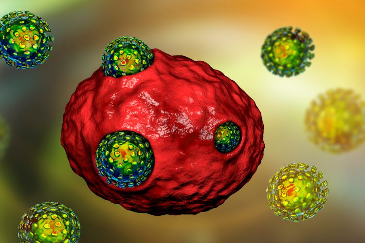
Lysosomes key to coronavirus shedding, finds study
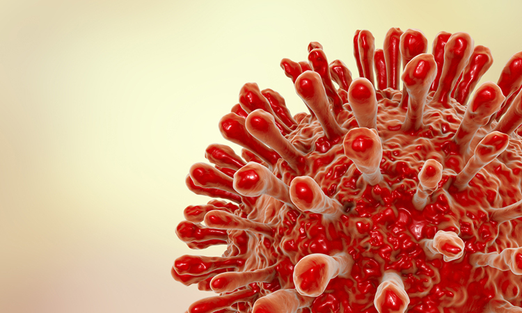
Scientists reveal that coronaviruses de-activate lysosomes before using them to exit infected cells and spread through the body.
List view / Grid view
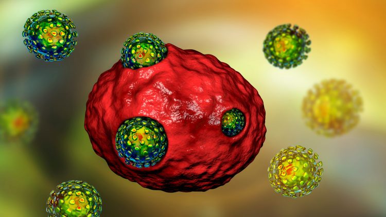
Scientists reveal that coronaviruses de-activate lysosomes before using them to exit infected cells and spread through the body.
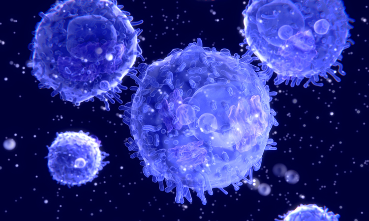
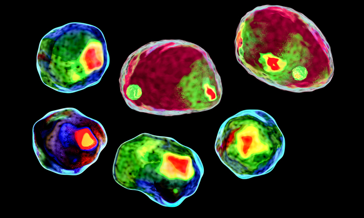
Researchers used flow cytometry to characterise which types of T cells are involved in the immune response to COVID-19 and what they target.
Identify therapeutic effects and adverse responses to compounds earlier in the drug discovery process.
Using cryo-electron microscopy and site-specific mass spectrometry, researchers have mapped the glycans that shield HIV from the immune system.
A new report has said that by 2023, the flow cytometry market will be worth $8.88 billion, partly due to an increase in stem cell research.
When delivered intranasally, the anti-inflammatory drug VX-765 prevented axon demyelination and loss in a murine model of multiple sclerosis (MS).
Combining 2D and 3D models with live-cell assays allows monitoring of cell responses in real time and provides important insights about compound treatment effects, biological complexity, and physiological relevance of assay results.

Learn how to perform complex analysis of calcium oscillations and assess cellular and mitochondrial toxicity.
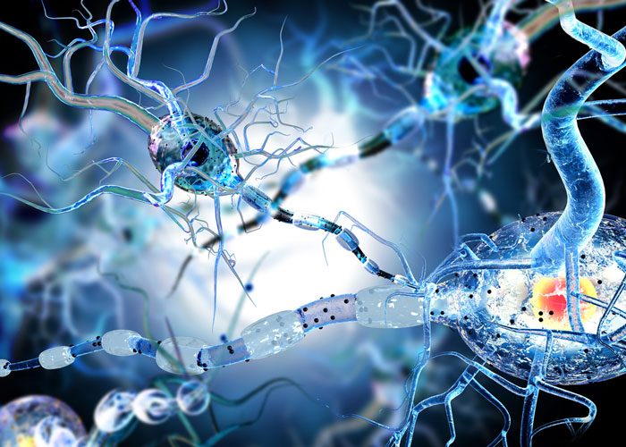
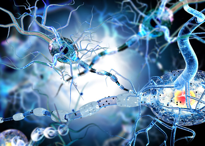
Neurological symptoms have been observed in COVID-19 patients. The link between SARS-CoV-2 and the CNS is critical to developing effective treatments

Water immersion objectives are essential for capturing more data at greater depths in 3D structures, such as spheroids and thick tissues.
Evaluating and quantitating nuclear transduction.
Researchers have found that the surface of SARS-CoV-2 can take on at least 10 different structural states when in contact with ACE2.

There has been growing interest in using high-content imaging methods for studying mitochondria.
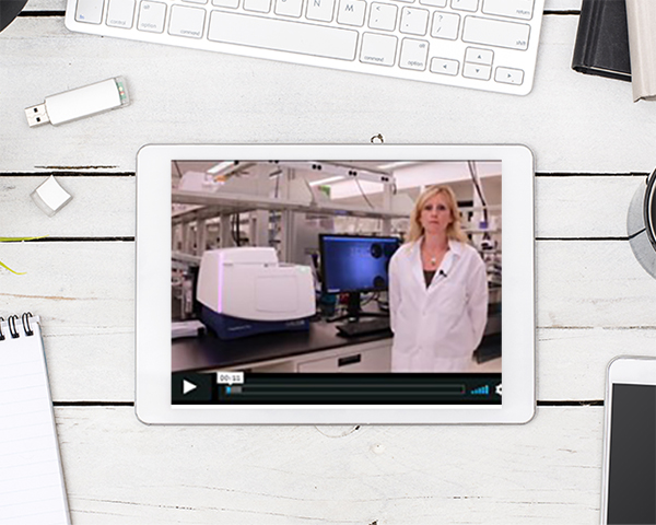
Explore this virtual tour of the ImageXpress Pico Automated Cell Imaging System from Molecular Devices, including hardware and software.
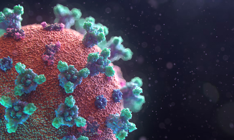
A new interactive map of the surface of SARS-CoV-2, featuring the Spike, Envelope and Membrane proteins, has been released for researchers to use.