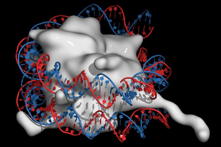
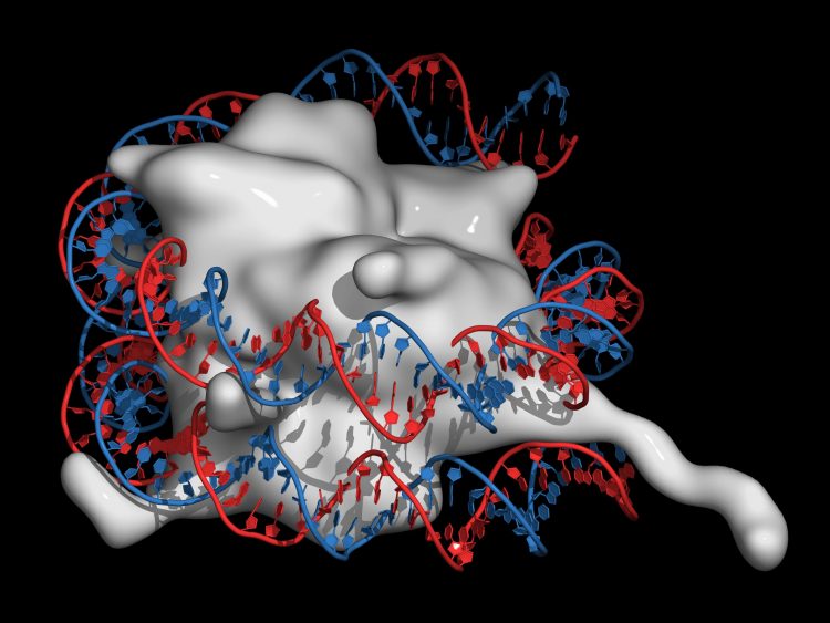
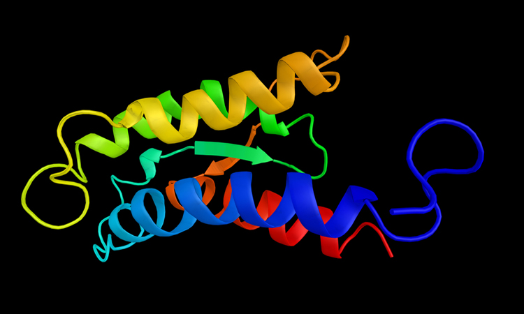
Interleukin-2 interactions revealed by researchers to aid in drug design
Using nuclear magnetic resonance spectroscopy, a study has shown that IL-2 can stimulate both effector T cells and regulatory T cells by adopting different structural forms.
List view / Grid view
Using nuclear magnetic resonance spectroscopy, a study has shown that IL-2 can stimulate both effector T cells and regulatory T cells by adopting different structural forms.
Researchers have created a new cryogenic electron microscopy (cryo-EM) technique by utilising low-energy electrons in a holographic method.
A new 3D model of the surface of the coronavirus COVID-19 has been released, to aid researchers in the development of a treatment.
Scientists in Hong Kong have developed a novel optical technique that facilitates accurate tracking of hemogenic endothelium cells in zebrafish embryos, providing new insights into the mechanisms of blood formation and potential new understanding of diseases such as leukaemia.
Researchers have developed a highly sensitive technique for probing molecules that they say could enable new applications in the fields of spectroscopy.
Researchers have identified that copper ions and their protein transporters, such as Atox1, are key to cancer cell movement and could be targeted by therapies.
Researchers have used virtual reality (VR) to control how drugs bind to their protein targets, which they say could be useful for designing new treatments.
Using fluorescent markers, researchers have developed Förster resonance energy transfer (FRET) to image the assembly, functions and interactions of molecules.
Researchers have shown how ATAD2, a histone chaperone protein, may load histones on to DNA in order to create the chromatin structure.
Scientists have identified two master controller regions that are essential for alpha-synuclein aggregation and could be targeted by future therapies.
Researchers have applied for a patent for their innovative cantilever and vibrating plate technique which they say could increase the speed of atomic force microscopy on fragile samples.
Researchers have used cryogenic electron microscopy to show that coronaviruses enter human cells through an interaction with angiotensin-converting enzyme 2 (ACE2).
A new imaging technique, which has revealed 3D forces exerted by tiny cell clusters, could help scientists understand how tissue forms, how wounds heal or how tumours spread.
The model was tested on a panel of drugs that are both still on the market or have been recalled due to adverse effects and was able to show their toxicity.
A new method to image cancerous tissues has been created by researchers who have paired infrared measurements with high-resolution optical images.