Stainless imaging developed by adding infrared to standard microscopes
Posted: 2 March 2020 | Victoria Rees (Drug Target Review) | No comments yet
A new method to image cancerous tissues has been created by researchers who have paired infrared measurements with high-resolution optical images.
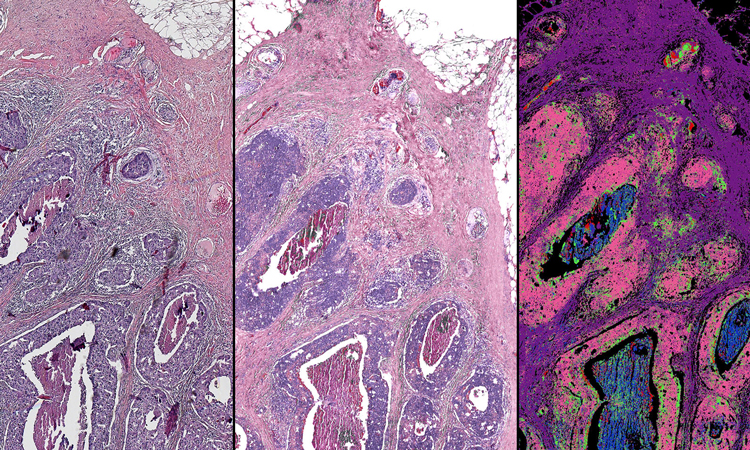
This side-by-side comparison of a breast tissue biopsy demonstrates some of the infrared-optical hybrid microscope’s capabilities. On the left, a tissue sample dyed by traditional methods. Centre, a computed stain created from infrared-optical hybrid imaging. Right, tissue types identified with infrared data. The pink in this image signifies malignant cancer (credit: Rohit Bhargava).
By adding infrared capability to the ubiquitous, standard optical microscope, researchers hope to improve cancer imaging for research and diagnosis. The team, from the University of Illinois at Urbana-Champaign, US, say they developed their stainless method to improve digital biopsies.
Pairing infrared measurements with high-resolution optical images and machine learning algorithms, the researchers created an imaging technique that closely correlates with traditional pathology processes and, according to the team, has outperformed state-of-the-art infrared microscopes.
“The advantage is that no stains are required and both the organisation of cells and their chemistry can be measured. Measuring the chemistry of tumour cells and their microenvironment can lead to better cancer diagnoses and better understanding of the disease,” said lead researcher Rohit Bhargava, a professor of bioengineering and the director of the Cancer Center at Illinois.
The scientists explain that when using a labelled technology, it can be difficult to distinguish cancer from healthy tissue or to pinpoint the boundaries of a tumour.
“For more than a century, we have relied on adding dyes to human tissue biopsies to diagnose tumours. However, the shape and colour induced by the dye provide very limited information about the underlying molecular changes that drive cancer,” Bhargava said.
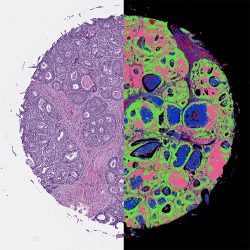
Machine-learning tools can analyse the data from the infrared-optical hybrid microscope to create digital versions of standard dyes, left, or to identify tissue types based on their chemical composition, right (credit: Rohit Bhargava).
Technologies like infrared microscopy can measure the molecular composition of tissue, providing quantitative measures that can distinguish cell types. Unfortunately, infrared microscopes are expensive and the samples require special preparation and handling, making them impractical for the vast majority of clinical and research settings.
Bhargava’s group developed its hybrid microscope by adding an infrared laser and a specialised microscope lens, called an interference objective, to an optical camera. The infrared-optical hybrid measures both infrared data and a high-resolution optical image with a light microscope – the kind ubiquitous in clinics and labs.
“We built the hybrid microscope from off-the-shelf components. This is important because it allows others to easily build their own microscope or upgrade an existing microscope,” said Martin Schnell, a postdoctoral fellow in Bhargava’s group and first author of the paper.
Combining the two techniques harnesses the strengths of both, the researchers said. It has the high resolution, large field-of-view and accessibility of an optical microscope. Furthermore, infrared data can be analysed computationally, without adding any dyes or stains that can damage tissues. Software can then recreate different stains or even overlap them to create a more complete, all-digital picture of the tissue.
“Infrared-optical hybrid microscopy is widely compatible with conventional microscopy in biomedical applications,” Schnell said. “We combine the ease of use and universal availability of optical microscopy with the wide palette of infrared molecular contrast and machine learning. And by doing so, we hope to change how we routinely handle, image and understand microscopic tissue structure.”
The researchers plan to continue refining the computational tools used to analyse the hybrid images. They are working to optimise machine-learning programmes that can measure multiple infrared wavelengths, creating label-free images that readily distinguish between multiple cell types and integrate data with the detailed optical images to precisely map cancer within a sample. They also plan to explore further applications for hybrid microscope imaging, such as forensics, polymer science and other biomedical applications.
The group published its results in the Proceedings of the National Academy of Sciences.
Related topics
Disease Research, Drug Targets, Imaging, Label-Free, Microscopy, Oncology, Research & Development
Related conditions
Cancer
Related organisations
Cancer Center at Illinois, University of Illinois at Urbana-Champaign
Related people
Rohit Bhargava