Novel deep learning tool developed to analyse microscopy images
Posted: 16 March 2021 | Victoria Rees (Drug Target Review) | No comments yet
An artificial intelligence platform has been created to enable tens of thousands of microscopy images to be generated in an hour.
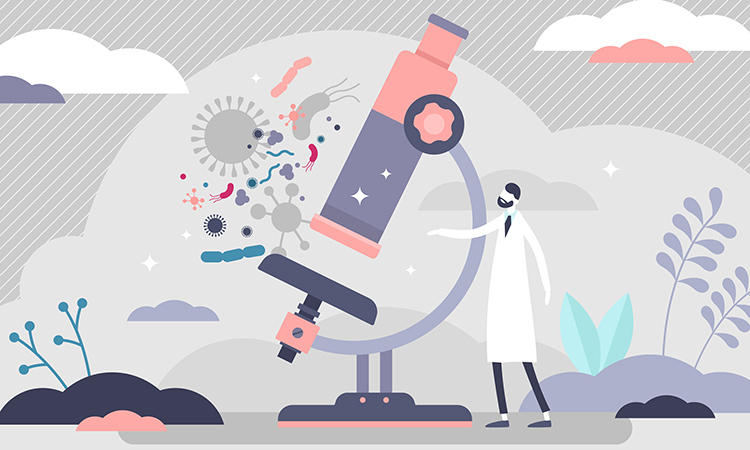
A novel artificial intelligence (AI) tool has been developed for analysing images taken with microscopes. A study shows that the tool can fundamentally change microscopy and pave the way for new discoveries and areas of use within both research and industry.
The technique, named DeepTrack 2.0, uses deep learning, a type of AI and machine learning. It was developed at the University of Gothenburg, Sweden.
“Deep learning has taken the world by storm and has had a huge impact on many industries, sectors and scientific fields. We have now developed a tool that makes it possible to utilise the incredible potential of deep learning, with focus on images taken with microscopes,” said Benjamin Midtvedt, a doctoral student in physics and the main author of the study.
The tool that the researchers developed involves neural networks learning to retrieve exactly the information scientists require from an image by looking through a huge number of images, known as training data. The tool simplifies the process of producing training data compared with having to do so manually, so that tens of thousands of microscopy images can be generated in an hour instead of a hundred in a month.
“This makes it possible to quickly extract more details from microscope images without needing to create a complicated analysis with traditional methods. In addition, the results are reproducible and customised, specific information can be retrieved for a specific purpose,” said Midtvedt.
The tool allows the user to decide the size and material characteristics for very small particles and to easily count and classify cells.
The researchers are hopeful that in the future DeepTrack 2.0 can be used to follow infections in a cell and map cellular defence mechanisms, which would open up huge possibilities for new medicines and treatments.
“We have already seen major international interest in the tool. Regardless of the microscopy challenges, researchers can now more easily conduct analyses, make new discoveries, implement ideas and break new ground within their fields,” said Midtvedt.
The study was published in Applied Physics Reviews.
Related topics
Artificial Intelligence, Disease Research, Imaging, Informatics, Microscopy
Related organisations
University of Gothenburg
Related people
Benjamin Midtvedt