Application note: Automated imaging of cell viability
Posted: 27 September 2019 | Cellular Imaging systems from Thermo Fisher Scientific | No comments yet
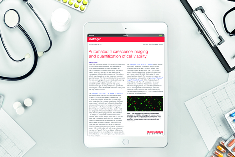
Automated fluorescence imaging and quantification of cell viability, with EVOS cell imaging system
Measuring cell viability is a key tool for studying cytotoxicity of compounds, infection, disease, and other aspects of cell health. While there are many cell viability and toxicity assays for high-throughput analysis, quantifying viability based on imaging of live and dead cells has typically been difficult and time-consuming. This makes it difficult to analyse many samples and obtain quantitative data.
With the recent introduction of automated fluorescence imaging systems, optimized viability staining kits, and powerful image analysis software, it is now possible to quickly and easily generate high-quality fluorescence images for many samples and quantify the percentage of live and dead cells to obtain cell viability data with high statistical power.
Related content from this organisation
Related topics
Assays, Drug Discovery Processes, Screening, Stem Cells, Targets, Translational Science
Related organisations
Cellular Imaging systems from Thermo Fisher Scientific