

Spatial proteomics in FFPE tumour tissue: an end-to-end workflow from prostate cancer research
Sponsored by  Miltenyi Biotec2026-10-28T14:00:002026-10-28T14:00:002026-10-28T15:00:00
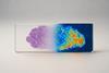
Follow a complete high-plex spatial proteomics workflow on archival FFPE tumour tissue, from marker-panel design to cell phenotyping, with a prostate cancer case study from the University of Surrey.
Archival formalin-fixed, paraffin-embedded (FFPE) tumour samples hold years of clinical history, but conventional profiling loses where cells sit in relation to each other. In the tumour microenvironment, where cells sit in relation to each other is part of the biology. High-plex spatial proteomics keeps that information, but turning the resulting high-dimensional data into a biological hypothesis is where many projects stall.
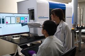
In this webinar, Dr Nicola Annels and James Thetford from the University of Surrey walk through an end-to-end spatial proteomics workflow on the MACSima Platform, step by step: marker-panel design, cyclic imaging, image quality control, cell segmentation and hierarchical phenotyping that separates tumour, stromal and immune compartments and identifies the major immune-cell subsets.
The case study is James Thetford’s PhD research into why prostate cancer outcomes differ between men of different ethnic and ancestral groups, and which features of the tumour microenvironment might contribute. It shows how tissue architecture, cellular composition, phenotypic states and spatial patterns can be analysed step by step to compare groups of patients, using a workflow that can be applied to archival FFPE tissue from other tumour types.
Attendees will also get practical guidance on designing spatial studies, interpreting the data, visualising high-dimensional results clearly and forming testable hypotheses from discovery data.
Access the free webinar by registering for a membership or logging into your account.