


International researchers have mapped the structure of the thromboxase A₂ receptor using cryo-electron microscopy, revealing some unexpected activation mechanisms.
Scientists have developed a detailed ’molecular map’ showing how a crucial human receptor involved in blood clotting and inflammation functions.
The study, led by an international team including researchers from Trinity College Dublin used advanced cryo-electron microscopy to capture high-resolution images of the thromboxane A₂ receptor while active and ready to transmit signals into cells.
Key role in blood clotting and disease
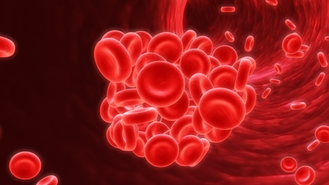
The thromboxane A₂ receptor is found on blood platelets and other cell types where it plays a central role in regulating clot formation, blood vessel contraction and inflammatory responses. Because of its wide-range of functions, it has long been a target of interest for scientists studying cardiovascular and inflammatory diseases.
The thromboxane A₂ receptor is found on blood platelets and other cell types where it plays a central role in regulating clot formation, blood vessel contraction and inflammatory responses
However, scientists have struggled to understand exactly how the receptor works, largely due to the fleeting nature of the molecule that activates it.
“Thromboxane A₂ itself is a short-lived signalling molecule that disappears within seconds in the body, which has always made it difficult for us to study how it activates the receptor,” said Dr Pawel Krawinski, Postdoctoral Research Fellow in Trinity College Dublin’s School of Medicine and School of Biochemistry and Immunology.
New technique unlocks structural detail
To overcome this challenge, the researchers applied an innovative imaging approach that allowed them to examine the receptor in extra detail. This helped them learn how it interacts with signalling proteins inside the cell.
The images that were captured presented several unexpected features. Unlike many similar receptors, the thromboxane receptor appears to rely on an unusual ’activation switch’ to trigger internal signalling. The team also found evidence that signalling molecules may enter the receptor from within the cell membrane rather than from outside the cell.
The researchers applied an innovative imaging approach that allowed them to examine the receptor in extra detail
“These insights are fascinating to us but they are far more than just structural details as they could have important medical implications too,” said Dr Krawinski. ”The thromboxane receptor plays a role in multiple diseases, including cardiovascular and cardiopulmonary disorders, pulmonary arterial hypertension and fibrotic lung disease, it is also overactive in some cancers and inflammatory conditions.”
The research also offers new understanding of rare inherited mutations affecting the receptor, which can lead to bleeding disorders. By examining how these genetic changes alter the receptor’s structure, scientists may be better equipped to diagnose and treat patients with such conditions.
Guiding future therapies
Overall, the study provides a new look at a clinically important signalling system. By combining structural biology, computational modelling and laboratory experiments, the researchers have created a comprehensive framework that could inform the development of safer and more effective therapies.
While further work is needed before these findings translate into clinical treatments, the molecular map represents an important step forward in understanding and targeting the thromboxane receptor in human disease.


No comments yet