To study the biological underpinnings of autism, researchers must examine the human brain itself. This article explores how Autism BrainNet supports this work through coordinated tissue donation and preservation.
Understanding autism at the level required for meaningful biological explanation depends on direct examination of the human brain. Imaging technologies, genetic sequencing and computational modelling have advanced neuroscience, but they cannot fully show how cells are organised, how circuits are altered or how molecular pathways operate within intact human tissue. For many research questions about neurodevelopmental conditions such as autism, postmortem brain tissue remains one of the few ways to obtain definitive answers.
For researchers studying autism and related conditions, brain donation is therefore a critical scientific resource for both basic neuroscience and early drug discovery. Direct access to human brain tissue allows investigators to identify disease-relevant cellular changes, validate molecular targets and map biological pathways that may be amenable to therapeutic intervention. Coordinating access to this material at scale is the role of Autism BrainNet, an international programme funded by the Simons Foundation and Simons Foundation International that collects, processes, stores and distributes donated postmortem brain tissue to support research worldwide.
My research focuses on understanding the cellular and molecular basis of neurodevelopmental disorders and on the development of antibody-based tools for neuroscience research.
One of the scientists working within this network is Dr Karl Murray. He is an adjunct professor in the Department of Physiology and Membrane Biology at the UC Davis School of Medicine and serves as a tissue coordinator at the University of California Davis MIND Institute node of Autism BrainNet.
“My research focuses on understanding the cellular and molecular basis of neurodevelopmental disorders and on the development of antibody-based tools for neuroscience research,” he explains. His role centres on receiving postmortem brain donations, processing them using protocols designed to preserve them for long-term research use and maintaining the collection so that samples can be distributed to approved investigators.
Autism BrainNet operates multiple collection sites across the United States and Canada, building a repository that enables scientists to examine autism at the cellular, molecular and structural levels in human brain tissue.
Why brain tissue remains irreplaceable
Although the human brain can now be studied through increasingly sophisticated non-invasive approaches, these technologies cannot replace the direct examination of tissue itself.
While there are many strategies for studying the human brain, such as magnetic resonance imaging (MRI) or electroencephalography (EEG), these methods do not allow direct access to the brain tissue.
“While there are many strategies for studying the human brain, such as magnetic resonance imaging (MRI) or electroencephalography (EEG), these methods do not allow direct access to the brain tissue,” Karl explains. “Brain tissue cannot be replicated in other ways, such as with artificial intelligence, computer models or virtual reality. Therefore, to study autism at the cellular and molecular level, access to brain tissue is essential.”
This access becomes particularly important when considering the diversity of autism itself. The condition encompasses a wide range of biological and behavioural profiles, meaning that large and well-characterised tissue collections are required to capture its full variability.
Autism BrainNet does not direct how research is conducted, but it evaluates requests for samples to ensure the best possible use of donated material. That care is critical, as a single brain donation can support hundreds of studies and remain viable for decades when processed correctly. To date, the collection contains more than 440 brain donations. Over half are from autistic individuals or from individuals with genetic conditions associated with high autism risk, alongside tissue from neurotypical controls.
Karl draws a parallel with another neurological field that has benefited enormously from access to postmortem tissue.
“There is so much to be learned from brain tissue, particularly if you consider how this type of work has advanced research in other neurological conditions, such as Alzheimer’s disease,” he says. “Scientists have been able to see the hallmark neurofibrillary tangles and plaques of Alzheimer’s Disease in postmortem brain tissue for more than a century. We now have biomarkers and treatments under development.”
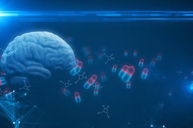
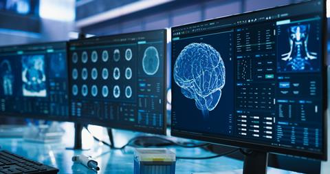
 Autism is a heterogeneous neurodevelopmental condition associated with differences in brain development and function. It is characterised by altered social communication, restricted or repetitive behaviours and atypical sensory processing. Co-occurring neurological or psychiatric conditions are common. Image credit: VisualMediaHub / Shutterstock.
[/caption]
Understanding autism biology at the cellular level
Autism is widely recognised as a neurological condition caused by genetic and environmental factors. Many individuals also experience co-occurring conditions such as epilepsy, anxiety, intellectual disability and sleep disorders, all of which relate to brain function.
Research using postmortem brain tissue provides one of the most direct paths to understanding the cellular changes that are associated with autism and related conditions.
“Research using postmortem brain tissue provides one of the most direct paths to understanding the cellular changes that are associated with autism and related conditions,” Karl says.
Such work is not focused on eliminating autism, he emphasises, but on deepening understanding and expanding options for individuals and families.
Improved biological understanding may eventually translate into better support, more tailored interventions or therapies that address specific biological pathways. It may also help explain why sensory processing differs between autistic and neurotypical individuals, or why certain co-occurring conditions develop in some people but not others.
The importance of rapid collection and careful preservation
Ensuring that donated brain tissue remains suitable for advanced research requires meticulous planning and rapid action. Cellular and molecular integrity begins to degrade after death, so timing is critical.
“To preserve the brain and maximise the viability of cellular and molecular components of the tissue, it needs to be collected within about 48 hours after death,” Karl explains.
For this reason, families are encouraged to consider donation in advance rather than making decisions during bereavement. Once donation is initiated through Autism BrainNet’s 24-hour hotline, trained professionals manage all of the logistics.
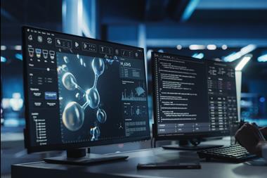
The preservation workflow is designed to maintain tissue integrity for a wide range of downstream analyses. Complementary preservation methods support both molecular studies, including genomic profiling and structural investigations that require intact tissue architecture. Each sample is carefully documented, with quality measures such as RNA integrity and genomic characterisation recorded to support reproducible research.
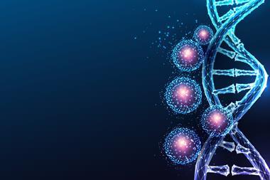
Researchers accessing the Autism BrainNet Tissue Catalogue can review specimen preparation protocols, postmortem intervals, RNA integrity measurements, phenotypic data and medical history. The organisation also records anecdotal social histories from families, providing contextual information about each donor’s life that may help interpret biological findings. In parallel, all donations from individuals with autism or related neurodevelopmental conditions undergo genetic characterisation using whole exome and whole genome sequencing.
These resources provide researchers with genetic and structural reference data that help link molecular risk factors to changes in brain biology, supporting target identification and mechanism-based therapeutic research.
Experimental approaches
The value of this extensive processing framework lies in the scientific flexibility it provides. Different preservation methods allow researchers to investigate distinct aspects of brain biology.
The ability to utilise this tissue for such a broad range of technical approaches underscores its utility to identifying molecular changes that associate with brain tissue in autism.
Fresh frozen tissue supports biochemical and genomic analyses, including protein isolation, nucleic acid extraction and modern single cell ‘omics’ techniques. Minimally fixed tissue retains spatial structure, enabling histopathological staining, inflammation markers and immunolabelling to map protein localisation.
“The ability to utilise this tissue for such a broad range of technical approaches underscores its utility to identifying molecular changes that associate with brain tissue in autism,” Karl explains.
By identifying molecular differences between autistic and neurotypical brains, researchers can begin to map biological pathways that may be relevant to symptoms and/ or therapeutic targeting.
What research has already revealed
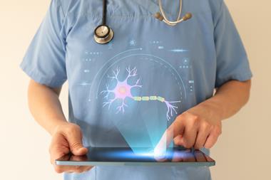
Postmortem studies have revealed measurable biological differences in autistic brain tissue, including altered molecular activity across systems involved in neuronal signalling, glial function and immune responses.
Earlier research typically analysed homogenised brain regions, providing bulk measures that averaged signals across many cell types. More recent approaches allow tissue to be dissociated into individual cells, enabling cell type specific analysis.
Single nucleus RNA sequencing studies, for example, have identified distinct molecular patterns across numerous neuronal and glial cell populations. Some of these changes appear to be driven by regulatory factors that control gene networks associated with autism risk.
Structural differences have also been reported. Quantitative analyses indicate altered architecture in certain brain regions, including reductions in inhibitory interneurons known as chandelier cells in the frontal cortex. These neurons play a key role in maintaining the balance between excitation and inhibition in neural circuits. Plus, their dysfunction may contribute to sensory sensitivity or co-occurring conditions such as epilepsy.
Questions for the future
Despite substantial progress, many scientific questions remain. Researchers are working to determine which biological changes are specific to autism and which are shared with other psychiatric conditions, including schizophrenia. Another major priority is understanding variability within the autistic population itself.
“There will also be findings that explain why some autistic individuals have co-occurring intellectual disability or anxiety and others do not,” Karl notes.
In allowing us to directly examine the changes that occur in the brain of individuals with autism, this donated tissue is incredibly valuable.
Over time, this knowledge may support the development of more targeted therapeutic approaches that address specific symptoms or underlying biological mechanisms.
“In allowing us to directly examine the changes that occur in the brain of individuals with autism, this donated tissue is incredibly valuable. Our hope is that it will inform our overall understanding of the neurobiology of autism which, in turn, will lead to a better understanding of its causes and improved quality of life for autistic individuals. From understanding of the basic biology of autism will come better lives for living autistic individuals in the future.”
Why access to human brain tissue matters
For early drug discovery and translational neuroscience, access to high quality human tissue remains a foundational resource. It enables the identification of molecular targets, validation of biological mechanisms and characterisation of disease heterogeneity – all essential for developing more precise therapeutic approaches.
Programmes such as Autism BrainNet depend on coordinated contributions from families, clinicians, tissue specialists and researchers to ensure donated material can be preserved, characterised and made available for scientific study.
For Karl and his colleagues, each donation provides direct access to human brain biology, supporting ongoing autism research and therapeutic development.
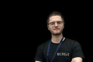
About the expert

Dr Karl Murray, Adjunct Professor at the University of California Davis School of Medicine
Dr Karl Murray, PhD, is an adjunct professor in the Department of Physiology and Membrane Biology in the University of California, Davis School of Medicine. As a trained neuroanatomist, Dr Murray’s research focusses on defining the impact of neurodevelopmental disease on mammalian brain architectural. In particular, Dr Murray is interested in how molecular and cellular heterogeneity define brain circuitry. Since 2025, he is also the Director of the UC Davis/NIH NeuroMab facility, focused on developing renewable, molecularly defined binders for proteomic studies in brain and he is also a tissue coordinator with Autism BrainNet. In this role, he is alerted to forthcoming brain donations and actively supports the collection, preservation and dissemination of heterogeneous, postmortem brain tissue for research.