Measuring disease progression remains one of the biggest hurdles in CNS drug development. Eye movements, now trackable with just a laptop and webcam, are emerging as a sensitive and scalable biomarker that could transform how trials are designed and therapies reach patients.
Quantifying progression: an urgent need in CNS drug development
Despite decades of intensive research, truly effective therapies for neurodegenerative diseases remain elusive. Conditions such as Parkinson’s disease (PD), amyotrophic lateral sclerosis (ALS), multiple sclerosis (MS) and Alzheimer’s disease (AD) continue to impose a growing and long-term burden on patients, caregivers and healthcare systems worldwide.1 The complexity of these disorders, which involve multiple pathways, diverse symptoms and highly variable clinical trajectories, makes drug development uniquely challenging.
One of the greatest obstacles lies in measuring how these diseases progress. Current ‘gold-standard’ scales, such as the Movement Disorder Society–Unified Parkinson’s Disease Rating Scale (MDS-UPDRS), ALS Functional Rating Scale–Revised (ALSFRS-R) and Expanded Disability Status Scale (EDSS) for multiple sclerosis, remain the dominant outcome measures.2 While well established, these tools are largely subjective and dependent on clinician observations and patient reports. This introduces variability between raters and even within the same rater over time, creating noise in clinical trial data.3 Moreover, these scales often miss the early or subtle changes that can be critical for determining whether a drug is effective.
Owing to these limitations, CNS drug trials often require large patient cohorts and long follow-up periods to achieve statistical power.4–6 This increases costs, slows timelines and ultimately delays the availability of treatments for patients. The pressing need is clear: the field requires objective, sensitive and scalable biomarkers that can more accurately monitor disease progression, improve trial efficiency and accelerate therapeutic development.
The quest for progression biomarkers
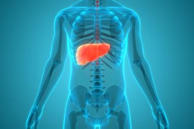
Over the past two decades, researchers have explored numerous biomarker approaches to address this challenge. Neuroimaging techniques, such as magnetic resonance imaging and positron emission tomography, have provided detailed insights into structural and functional changes in the brain. However, their high cost, limited accessibility and lack of scalability for large multicentre studies restrict widespread adoption.7

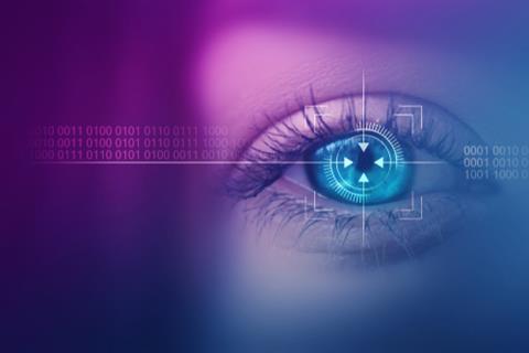
Neuroimaging methods such as MRI reveal structural and functional brain changes in diseases such as Parkinson’s, ALS, and MS. Yet their cost and limited scalability make them less practical as progression biomarkers in large clinical trials. Image credit: Gorodenkoff / Shutterstock[/caption]
Digital health tools, particularly wearables and smartwatches, have gained traction for their ability to monitor motor function and daily activity in real-world settings. Yet while promising, these devices often prove cumbersome and face long-term compliance challenges in patient populations already burdened with chronic disease.8
Fluid biomarkers, such as blood-based or cerebrospinal fluid measures, offer another route. However, these require invasive procedures and, in many cases, provide indirect measures of disease progression without clear correlations to functional outcomes.9
Against this backdrop, eye movements stand out as a particularly promising option. The neural control of eye movements involves widespread cortical and subcortical networks, meaning that abnormalities in oculometrics can serve as a sensitive reflection of brain dysfunction.10 Research has demonstrated that disruptions in saccadic latency, gain, velocity, fixation stability and intrusion frequency occur across conditions such as ALS, PD and MS.11–13
For years, however, the translation of this science into clinical practice was limited by the need for specialised, lab-based equipment,14 thus the arrival of AI-powered, video-based tools has changed this landscape dramatically. Using only a laptop and standard webcam, clinicians can now capture accurate and reliable oculometric data, making eye movements a truly practical candidate biomarker for clinical trials.15,16
Eye movements as an endpoint in clinical trials
The use of eye metrics has already moved beyond theory and into active drug studies, offering strong proof-of-concept for their utility.
- In a Phase II Parkinson’s trial, saccadic eye movements were used to monitor drug safety, demonstrating the feasibility of incorporating oculometric endpoints into clinical protocols17
- In spinal muscular atrophy, researchers assessed eye movements as part of drug efficacy evaluations, illustrating the adaptability of oculometrics even in populations with severe physical impairments18
- In a Phase IIb ALS trial, longitudinal results showed that the frequency of saccadic intrusions increased in parallel with disease progression.19 Importantly, these measures correlated with established clinical scales, further supporting their validity.20
- In another Parkinson’s study, progressive saccadic hypometria was detected over nine months, despite stable MDS-UPDRS III motor scores. Post-hoc analysis indicated that replacing the 21-month MDS-UPDRS endpoint with a nine-month eye movement endpoint could reduce required sample size per arm from 360 to just 140 participants.21
These studies underscore several advantages. Eye movement endpoints appear to be more sensitive to disease-related changes than traditional scales, offering earlier signals of therapeutic benefit. They are also highly practical: assessments take only about 10 minutes, require only a laptop and webcam, and can be repeated frequently. For patients and trials involving multiple sites, this means lower burden, easier logistics and richer datasets for analysis.
From innovation to integration: what’s next
Looking forwards, several trends are likely to accelerate the use of eye-based biomarkers. Continued improvements in AI algorithms and machine learning will strengthen accuracy and ease of deployment and the growing adoption of decentralised and hybrid trial models further supports integration, enabling frequent testing without compromising data quality.
Crucially, early evidence shows that eye movement metrics correlate strongly with established clinical scales. This means they can both complement and enhance traditional measures, offering a more detailed view of disease progression. As datasets expand, machine learning will likely uncover subtle, disease-specific signatures. This could allow researchers to stratify patients more precisely, monitor treatment response in real time and design trials that are shorter, smaller and more efficient.
Conclusion
Eye movements have emerged as a powerful and practical progression biomarker in CNS drug development. It is objective, sensitive, easy to implement and grounded in strong physiological mechanisms. By leveraging standard webcams and user-friendly interfaces, it offers a scalable solution suited for large, multicentre CNS drug trials.
When integrated into routine practice, eye movements could transform the clinical trial landscape; reducing sample sizes, shortening timelines and ultimately increasing the likelihood of bringing effective treatments to patients more quickly. For a field that has struggled for decades with slow and costly drug development, this represents a significant step forward.
Meet the Author
Eitan Raveh, Ph D, is Vice President of Clinical Partnerships at NeuraLight. A global clinical and regulatory affairs professional, he has more than 20 years of experience spanning medical centres, healthcare companies and the medical device industry. Eitan began his career in clinical neurology before earning his PhD from the Faculty of Medicine at Tel Aviv University, where he specialised in the application of advanced technologies in medicine. He has since held leadership roles across multiple medical and healthcare organisations, combining deep clinical expertise with a strong business perspective to drive the adoption of innovative medical technologies. He joined NeuraLight in 2022 to lead clinical partnerships and help advance the company’s biomarker platform in CNS drug development.
References
- Kumar D, Md Ashraf G, Bilgrami AL, Imtaiyaz Hassan M. Emerging therapeutic developments in neurodegenerative diseases: A clinical investigation. Drug Discovery Today 2022;27(10):103305. https://doi.org/10.1016/j.drudis.2022.06.005
- Regnault A, Boroojerdi B, Meunier J, et al. Does the MDS-UPDRS provide the precision to assess progression in early Parkinson’s disease? Learnings from the Parkinson’s progression marker initiative cohort. J Neurol 2019;266(8):1927–1936. https://doi.org/10.1007/s00415-019-09348-3
- Genge A, Cedarbaum JM, Shefner J, et al. The ALSFRS-R Summit: a global call to action on the use of the ALSFRS-R in ALS clinical trials. Amyotrophic Lateral Sclerosis and Frontotemporal Degeneration 2024;25(3–4):382–387. https://doi.org/10.1080/21678421.2024.2320880
- Delenclos M, Jones DR, McLean PJ, Uitti RJ. Biomarkers in Parkinson’s disease: Advances and strategies. Parkinsonism & Related Disorders 2016;22:S106–S110. https://doi.org/10.1016/j.parkreldis.2015.09.048
- Harris V, Tuddenham J, Sadiq S. Biomarkers of multiple sclerosis: current findings. DNND 2017;Volume 7:19–29. https://doi.org/10.2147/DNND.S98936
- Blennow K, Zetterberg H. Biomarkers for Alzheimer’s disease: current status and prospects for the future. J Intern Med 2018;284(6):643–663. https://doi.org/10.1111/joim.12816
- Brooks D. Assessment of neuroimaging techniques as biomarkers of the progression of Parkinson’s disease. Experimental Neurology 2003;184:68–79. https://doi.org/10.1016/j.expneurol.2003.08.008
- Hirczy S, Zabetian C, Lin Y-H. The current state of wearable device use in Parkinson’s disease: a survey of individuals with Parkinson’s. Front Digit Health 2024;6:1472691. https://doi.org/10.3389/fdgth.2024.1472691
- Irwin KE, Sheth U, Wong PC, Gendron TF. Fluid biomarkers for amyotrophic lateral sclerosis: a review. Mol Neurodegeneration 2024;19(1):9. https://doi.org/10.1186/s13024-023-00685-6
- Anderson TJ, MacAskill MR. Eye movements in patients with neurodegenerative disorders. Nat Rev Neurol 2013;9(2):74–85. https://doi.org/10.1038/nrneurol.2012.273
- Pretegiani E, Optican LM. Eye Movements in Parkinson’s Disease and Inherited Parkinsonian Syndromes. Front Neurol 2017;8:592. https://doi.org/10.3389/fneur.2017.00592
- Antoniades CA, Spering M. Eye movements in Parkinson’s disease: from neurophysiological mechanisms to diagnostic tools. Trends in Neurosciences 2024;47(1):71–83. https://doi.org/10.1016/j.tins.2023.11.001
- Donaghy C, Thurtell MJ, Pioro EP, et al. Eye movements in amyotrophic lateral sclerosis and its mimics: a review with illustrative cases. Journal of Neurology, Neurosurgery & Psychiatry 2011;82(1):110–116. https://doi.org/10.1136/jnnp.2010.212407
- Shaikh AG, Ghasia FF. Saccades in Parkinson’s disease: Hypometric, slow, and maladaptive. Progress in Brain Research. Elsevier p81–94. https://doi.org/10.1016/bs.pbr.2019.05.001
- Rosset I, Raveh E, Shimon AB, et al. Validation of a novel software‐based platform to extract oculometric measures. Acta Ophthalmologica 2022;100(S275):j.1755-3768.2022.0359. https://doi.org/10.1111/j.1755-3768.2022.0359
- Band TG, Bar-Or RZ, Ben-Ami E. Advancements in eye movement measurement technologies for assessing neurodegenerative diseases. Front Digit Health 2024;6:1423790. https://doi.org/10.3389/fdgth.2024.1423790
- Ellmerer P, Peball M, Carbone F, et al. Eye Tracking in Patients with Parkinson’s Disease Treated with Nabilone–Results of a Phase II, Placebo-Controlled, Double-Blind, Parallel-Group Pilot Study. Brain Sciences 2022;12(5):661. https://doi.org/10.3390/brainsci12050661
- Yae Y, Yuge K, Maeda T, et al. Exploratory evaluation of an eye-tracking system in patients with advanced spinal muscular atrophy type I receiving nusinersen. Front Neurol 2022;13:918255. https://doi.org/10.3389/fneur.2022.918255
- Berkman O, Raveh E, Harpaz E, et al. Changes in saccadic intrusions over time as an objective biomarker to follow ALS disease progression. Amyotrophic Lateral Sclerosis and Frontotemporal Degeneration 2024;25(7–8):760–766. https://doi.org/10.1080/21678421.2024.2376732
- Raveh E, Ben-Shimon A, Anisimov V, et al. Correlation between oculometric measures and clinical assessment in ALS patients participating in a phase IIb clinical drug trial. Amyotrophic Lateral Sclerosis and Frontotemporal Degeneration 2023;1–7. https://doi.org/10.1080/21678421.2023.2196315
- Gurevich T, Ezra A, Harpaz E, et al. Monitoring Parkinson’s Progression: Eye Movements vs. MDS-UPDRS III. Parkinsonism & Related Disorders 2025;134:107389. https://doi.org/10.1016/j.parkreldis.2025.107389
- Shaikh AG, Zee DS. Eye Movement Research in the Twenty-First Century—a Window to the Brain, Mind, and More. Cerebellum 2018;17(3):252–258. https://doi.org/10.1007/s12311-017-0910-5
Topics
- Analytical Techniques
- Artificial Intelligence (AI)
- Biomarkers
- Companies
- Disease Research
- Diseases
- Dr Eitan Raveh (Vice President of Clinical Partnerships at NeuraLight)
- Drug Development
- Drug Discovery Processes
- Imaging & Diagnostics
- Informatics
- NeuraLight
- Neurological disorders
- Rare & Genetic Disorders
- Translational Science