


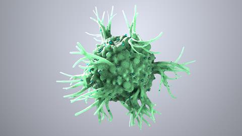
Researchers at the Icahn School of Medicine at Mount Sinai have pinpointed type 1 dendritic cells as the essential organisers of tertiary lymphoid structures within tumours, providing a mechanistic explanation for why TLS-rich tumours respond more favourably to immunotherapy.
Tumours that contain tertiary lymphoid structures (TLSs) are often associated with better patient outcomes and stronger responses to immunotherapy, but exactly how these immune hubs develop has been a question scientists haven’t yet managed to answer.
Now, researchers at the Icahn School of Medicine at Mount Sinai have published a new paper in Science, detailing a specialised subset of type 1 dendritic cells (DC1s) as the key organisers responsible for building and maintaining these structures. The study suggests these cells could become an important therapeutic target for boosting anti-tumour immunity.
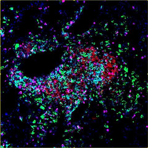
Using multiplex imaging, spatial transcriptomics and a newly developed mouse model that recapitulates human TLS formation, the team demonstrated that DC1s are required not only to initiate TLS development but also to sustain their function throughout tumour progression. Removing these cells disrupted the structures, while activating them enhanced local immune organisation.
Rather than acting only as antigen-presenting cells that activate T cells before migrating away, the researchers found DC1s remain embedded within tumours, orchestrating interactions between cytotoxic T cells, B cells and other immune populations to sustain local immune responses.
The findings give scientists crucial new information into why TLS-rich tumours respond more favourably to immunotherapy and suggest strategies aimed at increasing or activating DC1s could enhance responses in patients whose tumours currently lack these immune niches.
What actually are tertiary lymphoid structures?
Tertiary lymphoid structures (TLSs) are organised clusters of immune cells that develop in tissues experiencing chronic inflammation, including many solid tumours.
Unlike conventional lymph nodes, TLSs form directly within diseased tissue.
They typically contain:
- Dendritic cells.
- T lymphocytes.
- B lymphocytes.
- Specialised blood vessels that recruit additional immune cells.
Because immune activation occurs inside the tumour itself, TLSs are thought to generate faster and more sustained anti-tumour responses than immune activation occurring only in distant lymph nodes.
Across multiple tumour types – including lung, colorectal, melanoma and renal cancers – the presence of TLSs has consistently been associated with improved prognosis.
Why this matters
For years, TLSs have been viewed primarily as a prognostic biomarker. Multiple studies have linked their presence with improved survival and better responses to immune checkpoint inhibitors across several cancers, yet relatively little was understood about the biology controlling their formation.
This study moves the field beyond correlation by identifying a cellular mechanism controlling TLS development.
For drug discovery researchers, this shifts attention towards interventions that actively promote TLS formation, rather than simply using their presence as a predictor of treatment response.
If researchers can therapeutically increase functional DC1 populations within tumours, it may become possible to convert immunologically ’cold’ tumours into ’hot’ tumours that are more responsive to checkpoint blockade or other immunotherapies.
The bigger picture
Cancer immunotherapy has mainly focused on enhancing T-cell activity through checkpoint inhibition or engineered cell therapies.
However, evidence now suggests that durable anti-tumour immunity depends on coordinated interactions between multiple immune cell types.
TLSs function much like miniature lymph nodes embedded inside tumours, creating local environments where immune cells can:
- Activate tumour-specific T cells
- Support B-cell maturation and antibody production
- Generate sustained immune memory
- Coordinate ongoing anti-tumour responses.
This new work identifies DC1s as the cellular ’architects’ responsible for maintaining this organisation.
Rather than only presenting tumour antigens, these dendritic cells appear to provide the structural framework that allows multiple immune populations to communicate effectively within the tumour microenvironment.
What this means for early drug discovery
Several scientific developments could come from the information gained through this study:
New therapeutic targets
Instead of targeting tumour cells directly, researchers may seek molecules that promote recruitment, survival or activation of DC1s within tumours.
Combination therapies
Agents that stimulate TLS formation could potentially complement checkpoint inhibitors, cancer vaccines or cell therapies.
Patient stratification
The abundance and activity of DC1s could become biomarkers for predicting which patients are likely to benefit from immunotherapy.
Platform development
The new mouse model developed by the researchers may provide a valuable preclinical platform for testing therapies designed to manipulate TLS biology.
Questions that still remain
While the study identifies DC1s as essential organisers of TLSs, several questions remain before the findings can be translated clinically.
Researchers still need to determine:
- Which molecular signals recruit and maintain DC1s inside tumours
- Whether increasing DC1 numbers alone is sufficient to generate functional TLSs
- How tumour type influences TLS biology
- Whether therapeutically inducing TLSs will improve patient responses in clinical trials.
Understanding these mechanisms will be essential for designing therapies that reliably change the tumour microenvironment.







No comments yet