


Scientists at King’s College London have developed a scalable new way to study neural organoids, allowing researchers to track brain activity over time with greater consistency and precision.
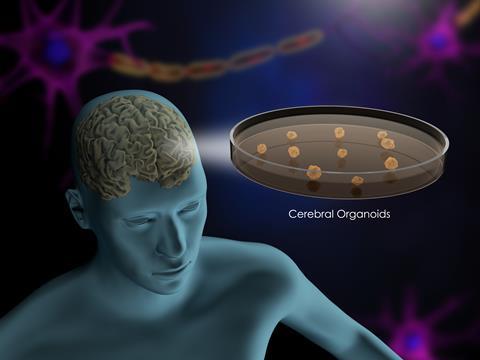
Neural organoids are a useful tool for understanding the human brain, giving researchers a crucial look into how brain tissue responds to drugs, how genetic mutations influence neural activity and how complex neural systems develop. Now, new research from King’s College London has moved closer to overcoming the longstanding limitations around scalability, reproducibility and longevity.
A breakthrough in organoid research
Scientists at King’s have developed an approach that enables neural organoids to be studied at greater scale and over longer periods. This development could change how researchers investigate neurological development, disease and potential treatments.
Functional genomic and pharmacological studies of neurodevelopment often depend on reliable measures of neuronal function, not just cell identity
“Functional genomic and pharmacological studies of neurodevelopment often depend on reliable measures of neuronal function, not just cell identity,” said Professor Deepak Srivastava, Professor of Molecular Neuroscience at King’s College London. “Our approach makes it possible to follow neural network activity over time and will allow us and others to directly compare the effects of drugs or gene variants across many parallel cultures.”
Challenges with existing models
Neural organoids grown in laboratories typically exist in either two-dimensional or three-dimensional forms, each with distinct advantages and disadvantages. Traditional 3D organoids closely resemble real brain tissue, containing a diverse mix of cell types. However, this diversity also introduces variability, making it difficult to reproduce results consistently, particularly in drug testing or gene studies.
Another major hurdle is the difficulty of recording electrical activity within 3D structures. Researchers are often limited to measuring activity at the surface or analysing individual neurons in isolation when probing deeper layers.
In contrast, 2D neural cultures allow scientists to track electrical signals across many neurons over time. Yet these simpler systems lack the rich cellular diversity found in 3D organoids and real brains, limiting their biological relevance.
Combining the best of both approaches
To address these issues, researchers including Dr Adam Pavlinek, Professor Anthony Vernon and Professor Deepak Srivastava developed a hybrid method. Their goal was to retain the cellular diversity of 3D organoids while gaining the scalability and accessibility of 2D systems.
The team began by growing 3D organoids before breaking them down into individual cells through a process known as dissociation.
The team began by growing 3D organoids before breaking them down into individual cells through a process known as dissociation. These cells, representing a wide range of developing neurons, were then cultured on flat surfaces. By pooling cells from multiple organoids, the researchers reduced variability and created more consistent neural networks.
This process resulted in multiple networks forming side by side on a single plate, all derived from similar cellular origins. The averaging effect of pooling helped smooth out differences between individual organoids.
Tracking brain activity over time
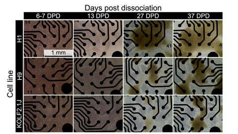
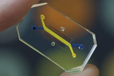
To monitor electrical activity, the cells were grown on microelectrode arrays, specialised plates embedded with electrodes. This allowed scientists to observe how neural networks formed and evolved over extended periods.
Unlike 3D organoids, which are difficult to study long-term, these 2D networks could be tracked for many days. Researchers observed neurons progressing from asynchronous activity, typical of early development, to synchronised firing patterns as connections matured.
Understanding variation in neural systems
The new approach also enables scientists to distinguish between technical and biological sources of variation. By placing similar cell populations alongside each other, researchers can directly compare how networks develop and respond to external influences such as drugs.
The hope is that this innovation can lead to more reliable and scalable models of the human brain
“The neurons in organoids have a remarkable ability of self-assembling into networks, we think the balance of neuron cell types in these networks may affect their electrical activity and may underlie the differences we see between networks,” said Dr Adam Pavlinek, first author.
The hope is that this innovation can lead to more reliable and scalable models of the human brain, opening new possibilities for research into neurological disorders and treatments.


No comments yet