
A blood vessel-on-a-chip model has enabled MIT researchers to programme capillary growth using controlled mechanical stretching, with PIEZO1 identified as a key molecular mediator – findings that could transform the fabrication of implantable engineered tissues.
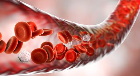
Researchers have developed a new technique to control the growth of blood vessels by mechanically stretching them, with the aim of overcoming one of the biggest challenges in engineering replacement tissues and organs.
Scientists have previously struggled to engineer living blood vessel tissue due to the intricacies of their networks, and without blood vessels to deliver oxygen and nutrients, engineered tissues cannot survive or function once implanted.
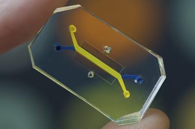
To combat this, engineers at the Massachusetts Institute of Technology (MIT) developed a human ’blood vessel on a chip’ capable of growing new capillaries in response to carefully controlled mechanical forces.
Stretching encourages blood vessel growth
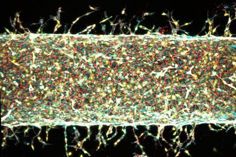
The MIT team developed a tiny laboratory model consisting of a central blood vessel formed from human endothelial cells embedded within a nutrient-rich gel containing a small magnet.
By using external magnets to repeatedly move the gel backwards and forwards, the researchers found they could stimulate the main vessel to produce new capillaries. The amount and direction of stretching also influenced how many vessels formed, how long they grew and the direction in which they developed.
The MIT team developed a tiny laboratory model consisting of a central blood vessel formed from human endothelial cells embedded within a nutrient-rich gel containing a small magnet
“Healthy tissues depend on organised blood vessel networks, but state-of-the-art protocols don’t enable fabricating such networks within engineered tissues,” says Ritu Raman, Associate Professor of Mechanical Engineering at MIT and the study’s co-lead author. “The ability to program blood vessel growth with physical cues may enable reproducible and scalable fabrication of engineered tissues that can be implanted in the body to restore function after debilitating disease or injury.”
When left undisturbed, the engineered artery produced only a small number of randomly positioned blood vessels. However, mechanical stretching significantly increased vessel growth. A five percent stretch produced the highest number of new capillaries, while stretching by 15 percent resulted in fewer but longer vessels.
New insight into how vessels respond
The researchers also discovered they could guide the direction of vessel growth by changing the direction of the stretching force, opening up the possibility of creating highly organised vascular networks for engineered tissues.
The researchers also discovered they could guide the direction of vessel growth by changing the direction of the stretching force
“The main takeaway is: stretching the blood vessel back and forth seems to enhance the number of new capillaries that grow,” Raman says. “We’re finding that moving is good, which is always the takeaway of everything we do in our lab. Mechanical forces play an important role in our bodies and that means that if you want to grow more or less vessels, or shorter or longer vessels, or vessels in certain directions, we now know how to do that.”
Gene identified as key regulator
To understand why blood vessels responded to mechanical stimulation, the team investigated the role of the PIEZO1 gene, which regulates ion channels that respond to physical pressure.
Using genetically modified endothelial cells in which the PIEZO1 gene had been suppressed, the researchers repeated the experiments and found that significantly fewer new blood vessels formed despite the same mechanical stretching.
This suggested that activation of PIEZO1 is a key part of the biological process linking physical forces to blood vessel growth.
The researchers now plan to use the technique to create organised vascular networks capable of supplying engineered organs and tissues with nutrients.


No comments yet