Scientists have developed an AI-enhanced imaging platform that enables non-invasive, label-free and longitudinal monitoring of cancer organoids and spheroids.

A team of European scientists have announced an artificial intelligence enhanced imaging platform that gives researchers a new way of studying cancer organoids and spheroids for non-invasive, label-free monitoring of tumour models over time.
Overcoming the limits of conventional imaging
Cancer organoids and spheroids have become key tools in cancer and pharmacological research. These three-dimensional models recapitulate tumour heterogeneity and key pathophysiological processes in vitro, while remaining relatively time and cost effective to generate.
Cancer organoids and spheroids have become key tools in cancer and pharmacological research.
However, most commercially available imaging systems rely on brightfield and fluorescence microscopy. While widely used, these approaches have limitations. They struggle to deliver non-invasive, high-content, label-free and longitudinal imaging simultaneously, especially when monitoring patient-derived organoids over extended periods.
To address this gap, researchers led by Professor Mengyang Liu at the Medical University of Vienna, Associate Professor Kristen Meiburger at the Politecnico di Torino and their collaborators developed an AI-enhanced optical coherence photoacoustic microscopy system known as OC-PAM.
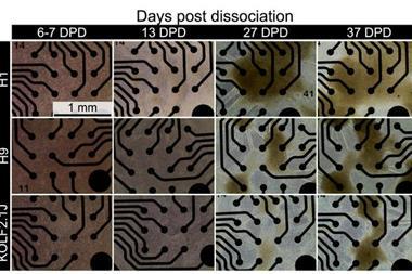
.jpg) Brest cancer organoids were imaged by OCM. Longitudinal tracking of organoids and viability evaluation of individual organoid were performed using the OCM data. Co-cultured breast cancer and melanoma spheroids were imaged by OC-PAM, with PAM signal being used to visualise the rare cell proxy. Credit: Mengyang Liu et al.[/caption]
Brest cancer organoids were imaged by OCM. Longitudinal tracking of organoids and viability evaluation of individual organoid were performed using the OCM data. Co-cultured breast cancer and melanoma spheroids were imaged by OC-PAM, with PAM signal being used to visualise the rare cell proxy. Credit: Mengyang Liu et al.[/caption] Tracking treatment response in real time
Through three carefully designed experiments, the team demonstrated that OC-PAM can perform longitudinal tracking of organoids, evaluate their response to chemotherapy, indicate individual organoid viability and identify proxies for drug tolerant persister cells. Crucially, all of these capabilities are achieved in a non-invasive and label-free way.
The findings highlight the system’s capacity to capture uncommon yet clinically significant behaviours within heterogeneous tumour populations.
For longitudinal imaging, the researchers employed the optical coherence microscopy mode to examine breast cancer organoids following chemotherapy exposure through carboplatin administration. Using automated tracking of individual organoids alongside quantitative analysis of their average volumes, the team assessed how the models responded to treatment.
Drug-treated organoids exhibited reduced growth rates. Notably, a small subset showed regrowth patterns consistent with drug-tolerant persister cells, rare cells believed to contribute to treatment resistance and relapse. This highlights the system’s capacity to capture uncommon yet clinically significant behaviours within heterogeneous tumour populations.
AI-driven insights into viability and rare cells
Beyond morphological changes, the study introduced a radiomics-based analysis of optical coherence microscopy data to evaluate organoid viability. By applying machine learning techniques, the researchers achieved high classification performance, showcasing the platform’s potential for non-destructive monitoring of treatment response over time.
In a further experiment, the team explored the system’s sensitivity to rare cell populations. Using OC-PAM, they imaged melanin-containing melanoma cells mixed with breast cancer cells within dense 3D spheroids. Even at very low concentrations, individual rare cells were successfully visualised.
Implications for cancer research and precision medicine
Together, the results show OC-PAM as a proficient system for studying cancer organoids and spheroids. By combining high-resolution structural imaging with AI-based analysis, the technology allows researchers to investigate drug resistance and rare cell populations in detail.
With its non-invasive, label-free and longitudinal capabilities, the system could help to advance cancer biology, accelerate drug development and support more personalised approaches to oncology.