


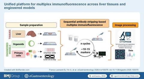
Researchers have developed a cost-effective, scalable multiplex immunofluorescence platform combining sequential antibody staining, conventional fluorescence microscopy and the open-source tool CytoPrixm to map cellular microenvironments in liver disease models.
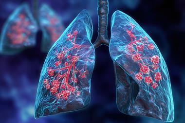
Spatial biology is allowing scientists to visualise how cells are organised and interact within intact tissues. In liver disease research, this is key because conditions including metabolic dysfunction-associated steatohepatitis (MASH), cholangiopathies, fibrosis and hepatocellular carcinoma are driven by complex and highly varied cellular environments.
Despite its potential, the wider adoption of multiplex imaging has been restricted by the high cost of specialist equipment and the complexity of data analysis. Now, researchers have developed an accessible multiplex immunofluorescence (mIF) workflow that uses standard laboratory equipment alongside open-source software, making advanced spatial analysis more widely available.
The study was led by Dr Marlene Kohlhepp, Dr Adrien Guillot and colleagues who created an integrated platform capable of analysing both liver tissue samples and engineered laboratory models.
Sequential imaging using standard laboratory equipment
The researchers based their approach on repeated cycles of antibody staining, imaging and antibody removal. After each round of imaging, antibodies were chemically stripped from the sample using a β-mercaptoethanol and SDS-based protocol, allowing new markers to be applied to the same tissue section.
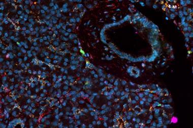
Using this method, the team successfully detected around 10 to 15 different markers within a single specimen while maintaining tissue architecture throughout multiple staining cycles. The workflow was specifically optimised for formalin-fixed paraffin-embedded (FFPE) liver tissue, the standard sample type used in pathology laboratories.
The technique enabled simultaneous visualisation of several key liver cell populations, including hepatocytes, cholangiocytes, macrophages, endothelial cells and hepatic stellate cells. This allowed researchers to build a detailed picture of the liver’s complex cellular microenvironment without requiring specialist imaging platforms.
Beyond tissue sections
The team also demonstrated that the workflow could be extended beyond conventional tissue samples to advanced laboratory models.
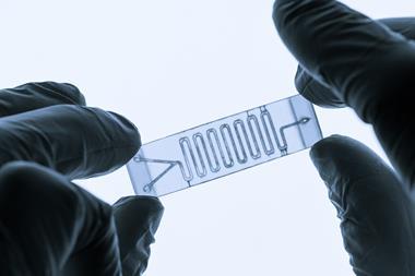
The protocol was successfully adapted for use with intrahepatic cholangiocyte organoids, primary liver cell cultures and liver-on-a-chip systems. Within organoids, researchers assessed epithelial polarity, cell proliferation and the integrity of cell-cell junctions by analysing markers including CK19, β-catenin, ZO-1, Ki67 and PCNA simultaneously.
In primary liver cell cultures, the platform enabled spatial mapping of hepatocytes, macrophages, endothelial cells and stellate cells grown together in the same environment.
Researchers also applied the workflow to a biliary niche-on-a-chip model containing multiple liver cell types, demonstrating its potential for studying disease mechanisms and evaluating new therapies in increasingly sophisticated experimental systems.
Open-source software simplifies analysis
To address the large volumes of imaging data produced by sequential multiplex analysis, the researchers developed CytoPrixm, an open-source image-processing software package.
The software combines several key preprocessing functions, including image stitching, background correction, channel alignment and DAPI-based image registration. By automating these processes, CytoPrixm reduces manual analysis and improves reproducibility while limiting the need for advanced programming skills.
The combination of accessible laboratory methods and freely available computational tools is designed to make spatial phenotyping practical for a wider range of research laboratories.
Supporting the future of precision hepatology
This platform could help improve translational liver research by enabling more detailed studies of immune cell recruitment, fibrogenesis, ductular reactions and tissue remodelling. It also provides a practical way to validate findings from emerging single-cell and spatial transcriptomics studies.
Rather than focusing on achieving the highest possible number of detectable markers, the workflow prioritises affordability, scalability and accessibility. By relying on commercially available reagents, conventional fluorescence microscopes and open-source software, the platform lowers many of the barriers that have previously limited the adoption of spatial biology technologies.
As spatially resolved molecular profiling becomes increasingly important in translational medicine, accessible imaging approaches like this could support advances in digital pathology, disease stratification and precision hepatology while broadening access to sophisticated spatial biology research.

No comments yet