


Why do some colorectal cancers resist immunotherapy? Analysis of patient tumour samples has identified a population of fibroblasts that could help explain treatment resistance.
Immune checkpoint blockade works by removing inhibitory signals that limit T-cell activity, allowing immune cells to recognise and attack tumour cells more effectively. However, treatment does not work for all patients, even when their tumours have molecular features associated with sensitivity.
Mismatch repair-deficient (dMMR) and microsatellite instability-high (MSI-H) colorectal cancers provide an important example. Defects in DNA mismatch repair allow mutations to accumulate, increasing the number of tumour-specific neoantigens that can be presented to and recognised by T cells. This high mutational burden can make these tumours particularly responsive to immune checkpoint blockade, yet a substantial proportion of patients still fail to respond.
Understanding this resistance requires looking beyond the tumour cells themselves. The surrounding tumour microenvironment contains immune cells, fibroblasts, extracellular matrix and signalling molecules that can influence whether cytotoxic T cells are able to reach and destroy cancer cells.
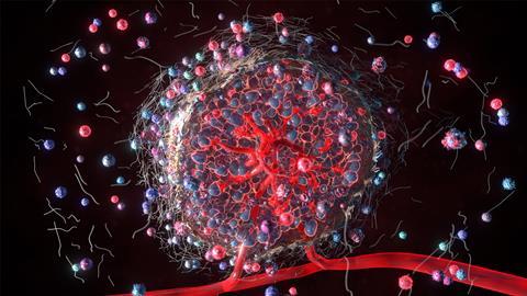
In a study published in Cancer Letters, researchers at Sun Yat-sen University Cancer Center analysed tumour samples from patients receiving immune checkpoint blockade to investigate differences between responders and non-responders. Using single-cell RNA sequencing and spatial transcriptomics, they identified a population of cancer-associated fibroblasts (CAFs) associated with treatment resistance. These cells accumulated around tumour-cell aggregates, contributing to a dense stromal barrier that restricted cytotoxic T-cell infiltration.
At a glance
The researchers investigated the tumour microenvironment of dMMR/MSI-H colorectal cancers to identify mechanisms associated with resistance to immune checkpoint blockade.
- 36 tumour samples from 34 patients analysed at single-cell level
- Responders compared with patients who did not respond to treatment
- Spatial analysis used to map cells within tumour tissue
- POSTN+ fibroblasts were more common in non-responders
- These fibroblasts formed dense regions that restricted T-cell access to tumour cells
- GDF15 signalling may contribute to their development
- Targeting the fibroblasts or surrounding matrix could provide a way to improve immunotherapy response.
Why some colorectal cancers resist immunotherapy
dMMR/MSI-H colorectal cancers are particularly immunogenic because defects in DNA mismatch repair allow mutations to accumulate. The resulting neoantigens can be recognised by T cells, helping to generate an anti-tumour immune response.
However, tumour genetics are only part of the picture. The tumour microenvironment can also determine whether immune cells are able to enter a tumour and remain functional. Fibroblasts are particularly important because they produce and remodel the extracellular matrix, which provides structural support to tissues but can also influence cell movement and signalling.
While stromal cells have previously been linked to T-cell exclusion in solid tumours, the fibroblast populations involved in immune exclusion in dMMR/MSI-H colorectal cancer have been less clearly understood.
Finding POSTN+ cancer-associated fibroblasts
Fibroblasts are cells that provide structural support to tissues, partly by producing collagen and other components of the extracellular matrix. Within tumours, fibroblasts can adopt altered states known collectively as cancer-associated fibroblasts (CAFs), which can influence tumour growth, immune activity and response to treatment.
Single-cell analysis identified several CAF populations within the tumour microenvironment, including a distinct group with high expression of periostin (POSTN), a protein involved in organising and remodelling the extracellular matrix. These POSTN+ CAFs were significantly more abundant in patients who did not respond to immune checkpoint blockade, suggesting a possible role in treatment resistance.
Further analysis showed that POSTN+ CAFs expressed genes involved in extracellular matrix organisation and collagen formation. Importantly, they were not evenly distributed throughout the tumour. Instead, they accumulated around groups of malignant cells, forming dense stromal regions and extensively remodelling the surrounding extracellular matrix.
These POSTN+ CAFs were significantly more abundant in patients who did not respond to immune checkpoint blockade, suggesting a possible role in treatment resistance.
Their location provided an important clue to their role in treatment resistance. The dense stromal regions appeared to restrict immune-cell access to malignant cells.
Keeping T cells outside the tumour
For immune checkpoint blockade to work effectively, cytotoxic T cells need to reach tumour cells and attack them. The researchers found that POSTN+ CAFs appeared to interfere with this process.
Spatial transcriptomic analysis showed that tumour-cell aggregates surrounded by POSTN+ CAFs contained significantly fewer infiltrating T cells, particularly CD8+ T cells. Multiplex immunofluorescence supported this finding, showing that in areas with high POSTN expression, CD8+ and CD4+ T cells tended to accumulate around the edges of these regions rather than penetrating the tumour core.
This suggests that the dense extracellular matrix produced by POSTN+ CAFs acts as a barrier between immune cells and malignant cells, limiting T-cell access to the tumour.
The fibroblasts may also affect T cells that become trapped within the surrounding stroma. Higher levels of POSTN+ CAFs were associated with greater T-cell exhaustion, a state in which prolonged stimulation reduces the ability of T cells to function effectively. In co-culture experiments, POSTN+ CAFs also reduced T-cell cytotoxicity more than POSTN-negative fibroblasts.
POSTN+ CAFs may therefore contribute to treatment resistance in two ways. They can restrict T-cell access to tumour cells and reduce the ability of nearby T cells to destroy them.
How do these fibroblasts develop?
The researchers next investigated how POSTN+ CAFs develop within the tumour. Rather than being a completely separate type of fibroblast, the findings suggest that they may arise as other fibroblasts change their molecular characteristics over time.
Their analysis suggested that fibroblasts expressing PI16 may transition to a state characterised by SFRP2 expression before developing into POSTN+ CAFs. This progression was seen predominantly in patients who did not respond to immune checkpoint blockade.
The researchers then looked at what might control this change. They identified increased activity of TWIST1 and TCF4, two transcription factors that regulate which genes are switched on or off within cells, as fibroblasts moved towards the POSTN+ state.
Experiments using primary fibroblasts isolated from human colorectal cancer tissue provided further evidence for their involvement, with both transcription factors promoting molecular characteristics associated with POSTN+ CAFs.
The findings suggest that resistant tumours may not simply contain more POSTN+ CAFs. Conditions within the tumour microenvironment may actively encourage other fibroblasts to develop into this treatment-associated state.
Tumour cells help create the barrier
The researchers explored what might drive fibroblasts towards the POSTN+ state. Their analysis identified growth differentiation factor 15 (GDF15), a signalling protein produced by tumour cells, as a potential factor.
GDF15 expression in malignant cells was higher in non-responders after treatment and was associated with higher POSTN expression in nearby fibroblasts. The researchers also found evidence of signalling between GDF15 and TGFBR2, a receptor on fibroblasts. When fibroblasts were exposed to GDF15 experimentally, they developed more characteristics associated with POSTN+ CAFs.
Tumour cells may therefore contribute to their own stromal barrier by signalling to nearby fibroblasts and encouraging them to adopt the POSTN+ state.
From human tissue to new therapeutic hypotheses
Only two patients provided matched samples from before and after treatment, and some of the proposed interactions between tumour cells and fibroblasts will require further experimental and in vivo validation before they can be considered established.
Even so, the study demonstrates the value of patient-derived tumour specimens for identifying mechanisms of treatment resistance that may not be apparent from tumour genetics alone.
Whether targeting POSTN+ CAFs or the stromal barrier they produce can improve response to immune checkpoint blockade remains an open question. However, the study provides a clear biological rationale for investigating the extracellular matrix, POSTN+ CAFs and GDF15 signalling as potential targets for overcoming resistance in dMMR/MSI-H colorectal cancer.





No comments yet