Vanitha Margan, Global Product Manager for Bio-Plex Multiplex Immunoassays at Bio-Rad Laboratories, reveals how multiplexing is being used to realise the full potential of extracellular vesicles in disease monitoring.
Extracellular vesicles (EVs) are emerging as powerful tools in the landscape of therapeutics research and diagnostics. These small, membrane-bound particles carry soluble analytes that reflect the state of their originating cells, making them powerful candidates for non-invasive biomarkers in many complex diseases like neurodegeneration, as well as innate immunity. However, EVs are highly heterogeneous – their molecular content varies depending on factors including source cell type, activation status, disease state, and environmental conditions, making single-marker approaches insufficient.1
Addressing the challenges of EV characterisation and realising their full potential for biomarker discovery demands the use of advanced technologies that can capture their dynamic and heterogeneous nature. The role of EVs in biomarker discovery can be transformed by enabling high-throughput, multi-analyte characterisation.
EVs as an alternative source of biomarkers
Biomarkers are fundamental indicators of disease state, progression, and response to therapy. Traditionally, tissue biopsies have been used for diagnostic and monitoring purposes in clinical studies to monitor these biomarkers. However, tissue biopsies can be invasive and often require repeated procedures to capture changes in disease over time. To address these limitations, researchers have turned their attention to EVs – a heterogeneous group of circulating vesicles that carry cargo, such as soluble proteins and nucleic acids, functioning as messengers between donor and recipient cells. Characterising these proteins and nucleic acids within EVs, including mRNA, microRNA, and DNA, offers minimally invasive insight into disease state and progression. Because EVs are highly enriched in a variety of bodily fluids, they are more suitable for repeat sampling compared to tissue biopsies, highlighting their diagnostic and prognostic relevance as well as their suitability for monitoring dynamic response to therapy (Figure 1).

Figure 1. EVs are found in most bodily fluids and can be captured through non-invasive liquid biopsy methods for downstream analysis of proteins and nucleic acid markers to support diagnosis and prognosis forecasting or to monitor response to therapy through changes in disease state and progression.[/caption]
The need for high-throughput, sensitive multiplex detection methods
Single-analyte assays, such as enzyme-linked immunosorbent assays (ELISAs), can detect individual biomarkers through enzyme-linked colorimetric, luminescent or fluorescent signals. However, their targeted approach cannot capture the complex biomolecular networks defining disease state and progression.2 Comprehensive profiling of multiple biomarkers would require numerous single-analyte assays to be carried out, demanding high sample volume and increasing the variability resulting from repeated sample handling and aliquoting. Furthermore, ELISAs are restricted by detection limits and cannot detect analytes present at very low concentrations.3 This is particularly challenging for EVs, whose protein and nucleic acid cargos can be found across a dynamic range of concentrations and are often scarce. These limitations demand a shift towards high-throughput, highly sensitive multiplexed approaches that use minimal sample volume to capture this complexity. This is an essential step towards realising the full potential of EVs for disease detection and treatment monitoring.
EV multiplex profiling strategies
EV multiplexing profiling strategies encompass technologies that enable the detection of multiple EV-derived analytes in a single assay, benefit from using smaller sample volumes, and can profile multiple analytes in a short period. These can be broadly categorised into two strategies: internal and external coding:
Internal coding
The first analytical approach relies on native molecular properties, such as charge/mass for high-throughput, mass spectrometry-based proteomic profiling.4 Mass spectrometry can give detailed molecular characterisation of biomolecules, including revealing post-translational modifications.5 However, as the diversity of biomolecules in a sample increases, it becomes more challenging to analyse the dynamic range of biomolecules present. To enhance detection sensitivity, abundant proteins can be removed or target proteins enriched.4 For example, immunodepletion can be used to capture albumin, endogenous immunoglobulins, transferrin, fibrinogen, and apolipoproteins present in plasma and the analytes of interest are recovered in the flow-through. Alternatively, proteins of interest can be enriched using ultracentrifugation or affinity chromatography. Mass spectrometry-based analysis enables precise biomolecular quantification, making it invaluable for research and biomarker discovery. However, its complexity and cost limit routine use in clinical settings, where faster and more practical methods are preferred.
External coding
External coding can take several forms: physical, which includes the use of capture arrays; chemical, which involves the use of chemical labels; biological, which uses nucleic acid labeling; and nanoparticle coding whereby nanoparticles with distinct optical or electrochemical properties are used as labelling tags for signal detection. These methods generally employ multiple receptors, including antibodies, aptamers, molecular beacons or locked nucleic acids, as well as multiple reporters to detect different analytes within the same sample.2 These approaches accelerate biomarker discovery in research and support efficient, multiplexed diagnostics in clinical practice.
Bead-based multiplex immunoassays
Among the most widely applied nanoparticle coding techniques are bead-based multiplex immunoassays. These use nano- or micrometer-sized colour-coded beads conjugated to a specific antibody to capture different analytes. To achieve colour-coding, the beads are created using two fluorescent dyes at distinct ratios to generate spectrally distinct signatures. When mixed with the sample, the addition of a detection antibody forms a sandwich complex, which is subsequently analysed using flow cytometry or similar technologies to provide quantitative data on the different analytes present (Figure 2). Some of the most commonly used biomarker research platforms are based on xMAP technology (x-multi analyte profiling, where ‘x’ represents the biomarkers), which relies on this approach and is capable of multiplexing up to 500 analytes in a reaction.6 Bead-based approaches can be adapted by conjugating different reagents to the beads, such as oligonucleotides, enzyme substrates, or receptors.

Figure 2. Bead-based multiplex immunoassays. A) Beads are internally coloured through two fluorescent dyes and bind to specific target analytes. Binding of detection antibodies conjugated to a reporter, eg, a biotinylated antibody, enables detection using reporter dyes. B) The use of multiple different beads enables multi-analyte detection in a single well.[/caption]
Preclinical and translational research applications of bead-based multiplexing
Bead-based multiplexing has emerged as a powerful approach for simultaneously detecting multiple analytes in EVs, offering significant advantages across multiple diseases.
In one observational study, urinary EV (uEV) presence was detected in COVID-19 patients during the early phases of kidney injury, suggesting their potential as early biomarkers for renal dysfunction.
In COVID-19, kidney injury is associated with disease severity and mortality, due to dysregulated inflammatory processes, such as cytokine storms. In one observational study, urinary EV (uEV) presence was detected in COVID-19 patients during the early phases of kidney injury, suggesting their potential as early biomarkers for renal dysfunction.7 The researchers also investigated whether the changes in the cargo carried by total and podocyte-derived uEVs were reflective of the inflammatory process during the early stages of kidney injury. Multiplex immunoassays enabled the simultaneous evaluation of multiple chemokines, cytokines and growth factors, revealing that the presence of urinary immune mediators within total uEVs predicted a higher risk of developing renal dysfunction. These findings highlight the ability of multiplexed EV profiling to identify at-risk patients and to capture the dynamics of organ-specific injury.
In Down syndrome (DS), much of the research has focused on the ageing trajectory of the broader DS population; however, less is known about the neurodevelopmental processes in infants and adolescents. Altered insulin signalling, and particularly its interplay with the mTOR pathway which plays a critical role in neuronal and glial differentiation, has been implicated in synaptic plasticity deficiencies, intellectual disability and neurodegeneration. Building on this insight, one study isolated neuronal-derived EVs (nEVs) from plasma samples of infants and adolescents with DS and applied multiplex immunoassay analysis to simultaneously evaluate mediators of the insulin/mTOR pathway to identify novel biomarkers and potential therapeutic targets.8 The results revealed significant pathway alterations, including IRS1 inhibition – a marker of brain insulin resistance associated with neuropathological alterations in DS. Comparable studies,9,10 have provided valuable information on molecular disruptions in Alzheimer’s disease, thus highlighting the diagnostic potential of nEVs and a minimally invasive approach for sampling neuronal components as well as the power of multiplexing to evaluate disruptions across entire signalling pathways quickly and efficiently.
Beyond EVs, multiplex immunoassays are also likely to revolutionise the potential of organoids for biomarker discovery and profiling in complex diseases.
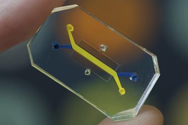
Beyond EVs, multiplex immunoassays are also likely to revolutionise the potential of organoids for biomarker discovery and profiling in complex diseases. Organ-on-chip technology can provide researchers with physiologically relevant 3D tissue models, offering a non-invasive alternative to tissue biopsies as well as more translationally relevant systems compared with animal models and 2D in vitro cultures. One recent study established a foetal- and maternal-derived organoid as a model for defining innate immune signalling at the maternal–foetal interface during pregnancy.11 The organoid model was used to investigate the antimicrobial contributions from foetal-derived placental trophoblasts and the maternal decidua during vertically transmitted viral infections. Matched trophoblast and decidua organoids were developed, and multiplexing was used to simultaneously profile the release of cytokines, chemokines and other factors at baseline and compare it to co-culture with human cytomegalovirus (HCMV). The findings revealed the unique antiviral signalling from both cells during infection. The study provides a strong foundation for the potential benefits of using organoids and multiplexing in research: firstly, that organoids are beneficial for studying immune signalling and secondly that multiplexing is well-suited for profiling multiple analytes using low sample volumes – an important factor when using organoids as sample volume is intrinsically low.
Collectively, these examples illustrate how bead-based multiplexed detection not only enhances biomarker discovery in EVs but also extends to innovative model systems such as organoids, broadening opportunities for translational research and precision medicine.
Conclusion
Soluble analytes serve as highly informative biomarkers in research and diagnostics. However, the complexities of many diseases make the analysis of a single biomarker ineffective and does not provide a full picture of the diverse and dynamic underlying disease biology. In research, preclinical, and clinical applications, where low sample volumes are favourable, EVs provide a minimally invasive resource for monitoring disease biology for research, diagnostic, and therapeutic purposes. The combination of EVs as minimally invasive biomarker sources and the analytical power of multiplexed detection represents a transformative strategy for understanding and monitoring complex diseases.
Moreover, the capabilities of multiplexing are transforming research using advanced in vitro models of disease, such as organoids, making it possible to tap into the full potential of these model systems to advance our understanding of the complexities of diseases.
References
- Kumar MA, Baba SK, Sadida HQ, et al. Extracellular vesicles as tools and targets in therapy for diseases. Signal Transduct Target Ther. 2024;9(1):27.
- Jiang C, Fu Y, Liu G, et al. Multiplexed profiling of extracellular vesicles for biomarker development. Nano-micro letters. 2021:14(1):3.
- Fang X, Yang Y, Wang H, Xu H. Bead-based microfluidic platforms for multiplex and ultrasensitive immunoassays in clinical diagnosis and treatment. Mechanobiol Med. 2024;2(2):100063.
- Van Gool A, Corrales F, Čolović M, et al. Analytical techniques for multiplex analysis of protein biomarkers. Expert Rev Proteomics. 2020;17(4):257-273.
- Whiteaker JR, Sharma K, Hoffman MA, et al. Targeted mass spectrometry-based assays enable multiplex quantification of receptor tyrosine kinase, MAP Kinase, and AKT signaling. Cell Rep Methods. 2021;1(3):100015.
- Pushparaj PN. Multiple analyte profiling (xMAP) technology coupled with functional bioinformatics strategies: potential applications in protein biomarker profiling in autoimmune inflammatory diseases. In: Shaik N, Hakeem K, Banaganapalli B, Elango R. (eds) Essentials of bioinformatics, volume II. Springer, Cham. 2019;151-165
- Medeiros T, Alves LS, Cabral-Castro MJ, et al. Exploring urinary extracellular vesicles and immune mediators as biomarkers of kidney injury in COVID-19 hospitalized patients. Diagnostics (Basel). 2022;12(11):2600.
- Perluigi M, Picca A, Montanari E, et al. Aberrant crosstalk between insulin signaling and mTOR in young Down syndrome individuals revealed by neuronal-derived extracellular vesicles. Alzheimers Dement. 2022;18(8):1498-1510
- Kapogiannis D, Mustapic M, Shardell MD, et al. Association of Extracellular Vesicle Biomarkers With Alzheimer Disease in the Baltimore Longitudinal Study of Aging. JAMA Neurol. 2019;76(11):1340-1351.
- Mullins RJ, Mustapic M, Goetzl EJ, Kapogiannis D. Exosomal biomarkers of brain insulin resistance associated with regional atrophy in Alzheimer’s disease. Hum Brain Mapp. 2017;38(4):1933-1940.
- Yang L, Semmes EC, Ovies C, et al. Innate immune signaling in trophoblast and decidua organoids defines differential antiviral defenses at the maternal-fetal interface. Elife. 2022;11:e79794.
Meet the expert
Vanitha Margan, Global Product Manager for Bio-Plex Multiplex Immunoassays, Bio-Rad Laboratories
 Vanitha Margan is Global Product Manager for Bio-Plex Multiplex Immunoassays at Bio-Rad Laboratories, where she leads global strategy, product lifecycle management, and customer education initiatives for Luminex xMAP-based multiplex immunoassays. With over 18 years of experience in clinical diagnostics and life sciences, her current work focuses on harnessing the power of multiplexing to help researchers unravel complex biological systems, driving meaningful discoveries in biomarker studies, and translational science.

Topics
- Analytical Techniques
- Assays
- Biomarkers
- Bio-Rad Laboratories
- Companies
- Disease Research
- Down syndrome (DS)
- Drug Targets
- Flow Cytometry
- Genomics & Sequencing
- High-Throughput Screening (HTS)
- Imaging & Diagnostics
- Immunoassays
- Immunology
- Infectious disease
- Informatics
- Kidney injury / renal dysfunction
- Mass Spectrometry
- microRNA
- Molecular Biology
- Neurological disorders
- Organoids
- Tools and techniques
- Translational Science
- Vanitha Margan (Global Product Manager for Bio-Plex Multiplex Immunoassays at Bio-Rad Laboratories)