


Many promising therapies fail because preclinical models do not fully capture the complexity of the human heart. A new review explores how 3D cardiac constructs could improve disease modelling, drug screening and safety assessment.
Cardiovascular disease remains a major cause of illness and death worldwide, but researchers continue to face challenges when evaluating potential treatments before they reach clinical trials. Animal models do not always reflect human biology, while traditional two-dimensional cell cultures lack many of the structural and functional characteristics of heart tissue.
In a review published in Research, scientists from Shanghai University and collaborating institutions suggest that 3D cardiac constructs could help address some of these shortcomings by providing more physiologically relevant models of human cardiac tissue.
The rise of 3D cardiac models
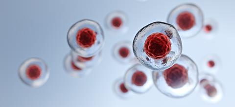
Researchers are developing a range of three-dimensional cardiac models designed to better replicate the structure and function of the human heart than conventional cell cultures. Most are generated from human pluripotent stem cells using approaches including scaffold-based systems, suspension cultures, microfluidic devices and self-organising protocols.
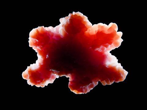
The review examines four of the main platforms currently used in cardiovascular research: cardiac organoids, engineered heart tissues, heart-on-chip systems and scaffold-free cardiac microtissues. Although all are designed to model heart biology, each has different strengths and applications.
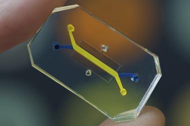
Engineered heart tissues are commonly used to assess contractile function, allowing researchers to measure how strongly heart muscle contracts in response to disease or drug treatment. Heart-on-chip platforms combine living cells with microfluidic technology to recreate aspects of the heart’s physical environment, including blood flow and mechanical stress.
Cardiac microtissues are better suited to larger-scale screening studies, making it possible to evaluate multiple compounds simultaneously. Cardiac organoids, meanwhile, can reproduce some of the structural and functional features of the developing heart, providing a way to investigate inherited disease and early stages of cardiac development.
Compared with conventional two-dimensional cell cultures, these models can better reproduce cardiac contraction, electrophysiology, metabolism and interactions between different cell types.
Capturing disease in the laboratory
Patient-derived induced pluripotent stem cells are becoming increasingly important in cardiovascular research because they allow scientists to create heart tissue carrying the same genetic variants found in patients. This makes it possible to study inherited disease in the laboratory using models that more closely reflect the biology of the people affected by it.
The review discusses the use of these cells to model inherited conditions including dilated cardiomyopathy, hypertrophic cardiomyopathy and arrhythmogenic cardiomyopathy.
Researchers can also recreate acquired disease states by exposing cardiac constructs to metabolic stress, inflammatory factors, environmental toxins or hypoxia. These approaches can be used to investigate processes such as oxidative stress, vascular dysfunction, inflammation and impaired contractility under controlled laboratory conditions.
Measuring cardiac function
One of the key challenges facing the field is ensuring that 3D cardiac models generate reliable and meaningful data. Researchers need to know not only whether a tissue resembles the heart structurally, but also whether it behaves like heart tissue.
To answer those questions, scientists measure features including electrical activity, calcium signalling, contraction strength, metabolism and gene expression. Techniques used include multielectrode arrays, patch-clamp recording, optical mapping, calcium imaging, extracellular flux analysis and multi-omics profiling. These measurements can reveal whether a drug alters cardiac rhythm, affects heart function or carries a risk of cardiotoxicity. However, many analytical methods were originally developed for two-dimensional cell cultures and may not be ideally suited to more complex 3D systems.
Biomaterials and AI
Improving the complexity of 3D cardiac models requires more than advances in stem cell biology alone. Researchers are also drawing on developments in biomaterials, bioengineering and artificial intelligence.
Conductive materials, hydrogels and 3D bioprinting techniques are helping to create tissue models that more closely resemble the structure and function of the human heart. At the same time, artificial intelligence is being used to analyse experimental data, classify cell types and support model design.
According to the review, AI-assisted approaches could help researchers identify promising compounds before they move on to more resource-intensive testing.
Remaining obstacles
Despite recent advances, several challenges continue to limit wider adoption.
Many stem cell-derived cardiomyocytes still resemble foetal or neonatal cells rather than mature adult heart cells, which can affect electrophysiology, metabolism, calcium handling and drug responses. Limited vascularisation restricts tissue size, long-term culture and cell survival, while reproducibility can be affected by differences in cell source, differentiation efficiency, culture conditions and assay design.
The review suggests that 3D cardiac constructs are unlikely to replace existing animal and in vitro models. Instead, they are expected to complement existing approaches by providing additional insight into disease mechanisms, drug efficacy and safety.
Wider adoption will depend on improvements in tissue maturation, vascularisation, standardisation and manufacturing, alongside clearer regulatory pathways for their use in drug development.


No comments yet