


The SEPARATE-Seq technique distinguishes tissue-infiltrating immune cells from circulating populations, revealing how the tumour microenvironment reshapes immune function and providing a clinically relevant framework for preclinical immunotherapy research.
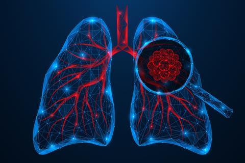
Researchers at Vlaams Instituut voor Biotechnologie (VIB) and Vrije Universiteit Brussel (VUB) have announced a new approach to studying how the immune system behaves within lung tumours, providing one of the most detailed immune maps yet of lung adenocarcinoma, the most common form of lung cancer. The findings combine a patient-relevant mouse model with advanced single-cell technologies to give researchers a new understanding of tumour biology.
A model that mirrors patient tumours
“We created a lung cancer model that closely mimics how tumours grow in patients,” says Professor Damya Laoui at VIB-VUB Center for Inflammation Research. “Combined with a new tracking method for cells, this allow us to tell the difference between immune cells inside the tumor tissue and those that are just passing by in the bloodstream. That distinction makes a big difference. It allows us to see much more clearly how immune cells behave and change once they are inside the tumour.”
We created a lung cancer model that closely mimics how tumours grow in patients
Lung cancer is the leading cause of cancer-related deaths worldwide, accounting for nearly 20 percent of all cancer deaths. Traditional preclinical research has largely relied on subcutaneous tumour models, where cancer cells are implanted under the skin. While these models are practical, they fail to replicate the lung’s unique immune environment.
To overcome this limitation, the team developed a lung adenocarcinoma model in which tumours grow directly within the lung. The model also allows tumour nodules to be separated from nearby healthy tissue, closely reflecting how clinical samples are handled. When compared with human datasets, the model successfully reproduced key immune characteristics seen in patients, including dysfunctional natural killer cells and increased regulatory and exhausted T cells.
“Our goal was to build a model that reflects what we actually see in patients,” says Pauline Bardet, PhD student at VIB-VUB and co-first author of the study. “By placing the tumour in its natural environment, the lung, we capture immune dynamics that are simply absent in subcutaneous models.”
Introducing SEPARATE-Seq
A key aspect of the study is a technique called SEPARATE-Seq, which stands for Streptavidin Enabled PARtitioning And Tag Evaluation for RNA-Sequencing. In complex organs such as the lung, immune cells are spread across blood vessels, tissue and airways, making it difficult to distinguish their precise location using standard methods.
“Location matters enormously,” explains Professor Damya Laoui, senior author of the study. “An immune cell inside a blood vessel is not experiencing the same signals as one embedded in tumour tissue. With SEPARATE-Seq, we can finally resolve that difference at single-cell resolution.”
By labelling immune cells in the bloodstream, SEPARATE-Seq enables researchers to identify which cells are actively infiltrating tumours and which are merely passing through. The method could also be applied to other diseases where immune cell positioning is critical.
Mapping immune activity inside tumours
Combining SEPARATE-Seq with spatial transcriptomics allowed the researchers to map not only the types of immune cells present but also their exact location within tumours. This revealed distinct spatial patterns, including a ring of lipid-associated macrophages around tumour edges and clusters of interferon-stimulated cells within tumours.
Combining SEPARATE-Seq with spatial transcriptomics allowed the researchers to map not only the types of immune cells present but also their exact location within tumours
The team also observed increased infiltration of hypoxic neutrophils, a higher presence of plasma cells and a shift in natural killer cells towards an immature and dysfunctional state after entering tumours. Many of these features were also identified in human samples, reinforcing the model’s clinical relevance.
“This level of spatial and molecular resolution allows us to see how immune cells specialise within defined tumour niches,” says Lize Allonsius, PhD student and co-first author of the study. “It highlights how strongly the tumour microenvironment reshapes immune function.”
A valuable resource for future research
Alongside the model, the researchers have released a comprehensive multiomics dataset accessible through an interactive online platform.
“Therapies succeed or fail based on how immune cells behave inside real tumours,” says Laoui. “If our models do not faithfully reflect patient biology, we risk drawing misleading conclusions. With this work, we provide a framework that brings preclinical research one step closer to the clinic.”


No comments yet