

Advances in organoid coculture – spanning Matrigel systems, air-liquid interface culture, microfluidics and bioprinting – are enabling researchers to recreate the complexity of the tumour microenvironment.
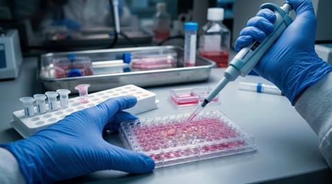
Three-dimensional cancer organoid models could provide more physiologically relevant platforms for studying tumour biology, evaluating immunotherapies and predicting drug responses, according to a new review examining advances in organoid coculture technologies.
Although cancer organoids grown in vitro can closely reproduce the genetic and phenotypic characteristics of their original tumour tissues, conventional culture methods often fail to capture the complexity of the tumour microenvironment (TME). This limits their ability to accurately represent how tumours interact with surrounding cells and respond to treatment.
Recreating the tumour microenvironment
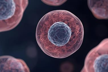
Cancer organoids are three-dimensional structures grown from tumour cells that retain important characteristics of the original tissue. They have become valuable tools for cancer research, drug screening and personalised medicine.
However, traditional organoid cultures typically lack several important components of the TME, including cancer-associated fibroblasts, immune cells and vascular networks. They also struggle to reproduce dynamic conditions found within tumours, such as fluid flow and oxygen gradients.
These limitations make it difficult to investigate interactions between tumour cells and surrounding tissues, model responses to immunotherapy and predict how patients might respond to particular drugs.
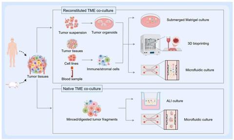
To address these challenges, researchers have developed several organoid coculture approaches designed to introduce additional TME components and create more representative models of tumour biology.
Four approaches to more representative cancer models
One approach involves submerged Matrigel coculture, which allows researchers to systematically introduce specific TME components into organoid cultures. This enables the investigation of defined interactions between tumour cells and other cell populations, helping researchers understand how individual components influence tumour behaviour.
Another strategy is air-liquid interface culture, which preserves the native tumour microenvironment. Unlike approaches that require the addition of selected cell populations, this technique retains resident immune cells and fibroblasts from the original tumour tissue. This makes it particularly useful for investigating immune cell infiltration and responses to immune checkpoint blockade.
Microfluidic culture systems and organoid-on-a-chip platforms provide greater control over the physical conditions surrounding tumour organoids. By introducing fluid flow, oxygen gradients and shear stress, these technologies overcome some of the limitations of conventional static cultures. They can also support the modelling of vascularisation and perfusion, alongside applications in high-throughput drug screening.
Meanwhile, three-dimensional bioprinting enables researchers to construct complex tumour architectures with precise spatial organisation. By positioning different cell populations within multilayered structures, researchers can recreate features of the TME, including vascular networks and interfaces between tumour cells and surrounding stromal tissue.
Applications in immunotherapy and personalised medicine
The review examines how these different coculture approaches could support the development and evaluation of cancer treatments.
More representative models could help researchers investigate interactions between tumour cells and immune populations, optimise immunotherapy strategies and assess adoptive cell therapies. They could also improve personalised medicine by providing experimental platforms that better reflect the biological characteristics of individual patients’ tumours.
However, each approach has its own advantages and limitations. While some offer greater control over individual cellular interactions, others prioritise the preservation of native tumour components or the recreation of complex physical environments.
The review compares these technologies and considers their suitability for different research applications, highlighting the need to balance biological complexity with experimental reproducibility and practical scalability.
Ultimately, continued development of organoid coculture systems could help address the limitations of conventional cancer models, providing more physiologically relevant preclinical platforms for investigating tumour biology and evaluating potential treatments.



No comments yet