

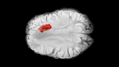
Researchers at KAIST and collaborating institutions have developed a patient-specific blood–brain tumour barrier chip that replicates the vascular environment surrounding glioblastoma tumours.
A laboratory-grown model that combines a patient’s glioblastoma cells with their surrounding blood vessel environment could offer drug developers a more realistic way to assess treatments before they reach clinical trials.
Developed by researchers at KAIST, Sungkyunkwan University, CHA Bundang Medical Center and CHA University, the patient-specific blood–brain tumour barrier (BBTB) chip is designed to reproduce one of the central challenges in treating glioblastoma: getting drugs through the brain’s vascular barrier and into the tumour.
The findings are particularly relevant to early drug discovery because they suggest that tumour genetics alone may not explain why apparently similar patients respond differently to the same treatment.
What is a BBTB?
The blood–brain tumour barrier is the altered vascular interface surrounding a brain tumour. It shares features with the normal blood–brain barrier but can become structurally and functionally different as a tumour develops.
For drug developers, this distinction matters because a promising compound may have potent activity against cancer cells in isolation but perform differently if it cannot reach those cells effectively within the tumour environment.
Why does this matter for early drug discovery?
Drug developers need laboratory models that can predict whether a candidate will work in humans. Yet conventional cancer models can struggle to reproduce the complex environment in which a drug must operate.
In glioblastoma, that environment includes the blood–brain barrier, which restricts substances moving from the bloodstream into the brain. The barrier is altered by the presence of a tumour but those changes can vary between patients.
Drug developers need laboratory models that can predict whether a candidate will work in humans
The new chip attempts to capture this complexity by combining patient-derived tumour cells with brain vascular endothelial cells and astrocytes. The system can also incorporate perivascular and immune cells.
That means researchers can assess more than whether a drug kills tumour cells. They can investigate whether it can cross a patient’s tumour-associated vascular barrier and how the surrounding environment influences its activity.
What did the researchers find?
The team created chips using tumour cells from three glioblastoma patients and tested temozolomide and bevacizumab, standard agents used in glioblastoma treatment.
All three patients had the same result for the MGMT promoter methylation biomarker, which would normally suggest broadly similar treatment responses.
The chips nevertheless showed differences in both the vascular barrier characteristics and drug responses of the three patient-derived models. The researchers reported that these findings showed a high level of agreement with the patients’ actual clinical courses.
This indicates the biological context surrounding a tumour may be as important to treatment response as characteristics within the cancer cells themselves.

How does this fit with the field?
The work sits within the rapidly developing field of patient-derived models and organ-on-chip technology.
Researchers are now trying to move away from simplified cell cultures towards models that reproduce interactions between tumour cells, blood vessels, immune cells and surrounding tissue. Patient-derived organoids and other three-dimensional systems can preserve some of the biological characteristics of individual cancers, while microfluidic platforms can introduce controlled fluid flow and tissue interfaces.
The work sits within the rapidly developing field of patient-derived models and organ-on-chip technology
The BBTB chip adds another layer by focusing specifically on the vascular barrier that drugs must negotiate in the brain.
It is not yet clear whether the model will outperform existing approaches in predicting clinical outcomes, as current study involved tumour material from only three patients, larger validation studies will be required.
What does it mean for researchers?
For academic researchers and drug developers, the platform could provide a way to investigate why the same compound behaves differently across patients.
It could also allow several compounds or treatment combinations to be compared using tumour cells from the same individual. In early development, this could support researchers in indentifying promising candidates and investigating potential mechanisms of treatment resistance before committing to more resource-intensive studies.
The model may also be useful for studying the barrier itself. Researchers could examine how new drug candidates interact with the tumour-associated vasculature and whether differences in barrier properties affect drug exposure.
“This study is meaningful in that it presents a platform that recreates patient-derived tumour cells together with the blood–brain tumour barrier, allowing patient-to-patient differences in treatment response to be evaluated in a way that closely reflects reality,” said Professor Song Ih Ahn, Assistant Professor in the Department of Medical Engineering at KAIST. “We hope to validate it in a larger patient population and develop it into a preclinical evaluation platform for establishing personalised treatment strategies and for new drug development.”
Key takeaway
The significance of the work is not simply that researchers have created another glioblastoma model. It is that the model attempts to reproduce the patient’s tumour and the vascular barrier surrounding it as a connected system.
If validated, that could give researchers a more informative tool for early drug development, particularly for understanding why promising treatments fail to work equally well across patients.
For now, however, the chip remains a preclinical research platform, not a clinically validated test for selecting treatments. The larger studies that follow will determine whether its promise translates into a practical drug development tool.
What happens next?
The immediate priority is validation, as the researchers plan to test the platform in a larger patient population to establish whether its apparent ability to reproduce treatment responses holds across a broader range of glioblastomas.
Further research will also need to establish whether the platform can distinguish between multiple investigational drugs and help identify effective combination treatments. Ultimately, the goal is to discover if the technology can develop into a routine preclinical model for evaluating new brain cancer therapies.





No comments yet