


The right signals can make or break an organoid model. From growth and differentiation to downstream drug testing, cytokines and growth factors are critical to creating cultures researchers can rely on.
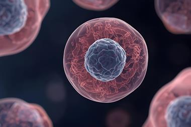
Organoids are three-dimensional, self-organising models derived from stem cells that recapitulate key structural and functional features of in vivo tissues. Cytokines and growth factors provide essential niche signals that regulate stem cell proliferation, self-renewal and differentiation.¹,² Classical factors such as R-spondin 1 (RSPO1), EGF, Noggin and Wnt3a – together with tissue-specific and emerging signals such as FGFs, NRG1 and IGF2 – support organoid establishment, expansion and differentiation.³,⁴
With growing applications in disease modelling, drug screening and precision medicine, reliable cytokines are increasingly important for reproducible organoid culture.⁴ Sino Biological provides high-quality cytokines with validated bioactivity and consistent performance to support diverse organoid culture and research applications.
Cytokine signalling in organoid culture
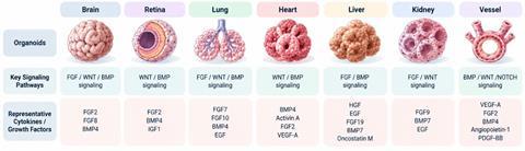
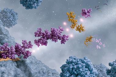
Organoid culture depends on the reconstruction of signalling environments that normally govern cell fate within tissues. Rather than acting independently, cytokines and growth factors form interconnected signalling networks that balance stem cell maintenance, proliferation and differentiation. A classic example is the WENR system, in which Wnt3a and RSPO1 reinforce Wnt/β-catenin signalling, EGF promotes epithelial proliferation and Noggin suppresses BMP signalling to help maintain the stem/progenitor state.⁵,⁶
Beyond this core framework, tissue- and stage-specific signals further shape lineage commitment, maturation and cellular composition. Recent organoid systems increasingly incorporate additional FGF, TGF-β/BMP, Notch and other pathways to reproduce more complex tissue patterning and cellular interactions.⁷ The composition and timing of these signals can therefore profoundly influence organoid identity and phenotype, enabling the development of models that more closely reflect the biology of their tissues of origin.
From cytokine-guided culture to translational applications
The ability to precisely control cytokine and growth factor signalling has expanded organoids from experimental 3D cultures into increasingly relevant models for translational research. Defined culture conditions support the establishment and expansion of tissue- and patient-derived organoids while helping to preserve key cellular, molecular and functional features of their tissues of origin.⁸,⁹
These models provide biologically relevant platforms for disease modelling, drug efficacy and toxicity testing, and preclinical therapeutic development.⁸ Patient-derived organoids further capture inter-patient heterogeneity and can be used to evaluate individualised treatment responses, supporting their growing potential in precision medicine.⁹,¹⁰ Importantly, reproducible cytokine and growth factor conditions are essential for generating consistent organoid phenotypes and reliable downstream readouts.¹¹
| Application | What organoids offer | Role of cytokines & growth factors |
|---|---|---|
|
Disease modelling |
Mimic tissue structure and function, Reproduce key disease features |
Maintain tissue-specific cell types, Support disease-related phenotypes |
|
Drug screening |
Test drug efficacy, Evaluate drug toxicity |
Support stable organoid growth, Improve experimental consistency |
|
Cancer research |
Model patient tumours, Preserve tumour heterogeneity |
Support tumour organoid growth, Maintain tumour cell diversity |
|
Precision medicine |
Compare treatment options, Predict individual drug responses |
Maintain patient-derived organoids, Support reliable drug testing |
|
Regenerative medicine |
Expand tissue-specific cells, Support tissue repair research |
Promote cell expansion, Guide differentiation and maturation |
|
Developmental research |
Model tissue formation and development, Study changes in cell fate |
Recreate developmental signals, Guide tissue-specific differentiation |
Applications in research
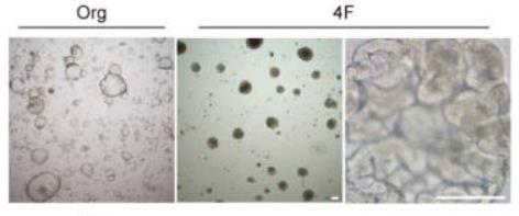
Recombinant growth factors are widely used to establish and maintain organoid models across different tissue types. For example, EGF and bFGF have been incorporated into culture systems for the generation of mouse pancreatic ductal organoids (Figure 2), providing an ex vivo model for studying pancreatic epithelial biology.
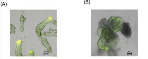
Similarly, R-Spondin has been used as a key component of mouse colonic organoid culture, supporting the transition from isolated colonic crypts to established organoids (Figure 3). These examples illustrate how defined growth factors can support organoid-based studies across diverse tissue models.
Sino Biological solutions for organoid research
Sino Biological provides a comprehensive portfolio of cytokines and growth factors to support organoid establishment, expansion, differentiation and functional research across diverse organoid models. Its organoid research solutions cover 3D organoid culture, physiological organoids and tumour organoids, providing researchers with relevant signalling factors for different tissue types, culture stages and research applications.
| Featured cytokine / growth factor | Cat. No. | Key features |
|---|---|---|
|
EGF |
>95% SDS-PAGE · Tag-free · Active |
|
|
Noggin / NOG |
>85% SDS-PAGE · hFc Tag · Active |
|
|
R-Spondin 1 / RSPO1 |
≥95% SEC-HPLC · Native · Active |
|
|
FGF2 / bFGF |
≥95% SDS-PAGE · Native · Active |
|
|
FGF10 |
≥95% SEC-HPLC · Native · Active |
|
|
Activin A |
≥95% SEC-HPLC · Native · Active |
|
|
IL-22 |
>95% SDS-PAGE · Native · Active |
|
|
VEGF165 |
≥95% SEC-HPLC · Native · Active |
|
|
TGF-β1 |
>95% SDS-PAGE · Native · Active |
|
|
IL-13 |
≥95% SEC-HPLC · Native · Active |
|
|
FGF4 |
≥95% SEC-HPLC · Native · Active |
|
|
β-NGF |
≥90% SEC-HPLC · Native · Active |
References
1. Zhao Z, Chen X, Dowbaj AM, et al. Organoids. Nature Reviews Methods Primers 2, 94 (2022). doi:10.1038/s43586-022-00174-y.
2. Clevers H. Modeling Development and Disease with Organoids. Cell 165, 1586–1597 (2016). doi: 10.1016/j.cell.2016.05.082.
3. Hendriks D, Artegiani B, Margaritis T, et al. Mapping of mitogen and metabolic sensitivity in organoids defines requirements for human hepatocyte growth. Nature Communications 15, 4034 (2024). doi:10.1038/s41467-024-48550-4.
4. Wu H, Liu S, Chen S, et al. Generation of proliferative hESC-derived grape-clustered hepatocyte organoids with multipolar architecture as regenerative counterpart via synergy of YAP and IGF2 pathways. Cell Death & Disease 17, 381 (2026). doi:10.1038/s41419-026-08635-y.
5. Sato T, Vries RG, Snippert HJ, et al. Single Lgr5 stem cells build crypt-villus structures in vitro without a mesenchymal niche. Nature 459, 262–265 (2009). doi:10.1038/nature07935.
6. Sato T, Stange DE, Ferrante M, et al. Long-term expansion of epithelial organoids from human colon, adenoma, adenocarcinoma, and Barrett’s epithelium. Gastroenterology 141, 1762–1772 (2011). doi: 10.1053/j.gastro.2011.07.050.
7. Xu M, Kong D, Sun S, et al. Organoids for disease modeling and treatment: state-of-the-art. Experimental Hematology & Oncology 15, 10 (2026). doi:10.1186/s40164-026-00743-x.
8. Tang XY, Wu S, Wang D, et al. Human organoids in basic research and clinical applications. Signal Transduction and Targeted Therapy 7, 168 (2022). doi:10.1038/s41392-022-01024-9.
9. Thorel L, Perréard M, Florent R, et al. Patient-derived tumor organoids: a new avenue for preclinical research and precision medicine in oncology. Experimental & Molecular Medicine 56, 1531–1551 (2024). doi:10.1038/s12276-024-01272-5.
10. Wensink GE, Elias SG, Mullenders J, et al. Patient-derived organoids as a predictive biomarker for treatment response in cancer patients. npj Precision Oncology 5, 30 (2021). doi:10.1038/s41698-021-00168-1.
11. Jin Y, Park S, Cho SW. Organoid analytical toolkits. Nature Reviews Bioengineering 4, 336–352 (2026). doi:10.1038/s44222-025-00384-5.









No comments yet