


Promising compounds don’t always engage their targets in living cells. At ELRIG Drug Discovery 2026, Dr Joshua Sopp of Promega discusses why earlier cellular testing matters and how BRETSA could help researchers investigate new drug targets.
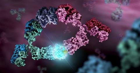
A compound may bind strongly to a purified protein, but that does not guarantee it will engage the same target inside a living cell. Protein interactions, post-translational modifications and compound permeability can all affect its behaviour, making cellular target engagement an important consideration in early drug discovery.
At ELRIG Drug Discovery 2026, Drug Target Review spoke with Dr Joshua Sopp, Product Specialist at Promega, about the limitations of existing assays and why measuring target engagement earlier could help researchers make better decisions about which compounds to take forward.
The discussion also covered Promega’s TarSeerTM BRETSATM Target Engagement System, which measures changes in protein thermal stability in living cells. By removing the need for existing target-specific chemical probes or validated antibodies, the technology could make it easier to investigate poorly characterised targets.
Why cellular target engagement matters
Confirming that a compound interacts with its intended target is an important step in early drug discovery. Biochemical techniques such as surface plasmon resonance (SPR) and thermal shift assays can provide evidence of molecular interactions using purified proteins.
However, these experiments do not necessarily reproduce the conditions in which a target operates inside a living cell.
Sopp explained that biochemical assays cannot always capture the conditions in which proteins naturally function, particularly when researchers need to work with purified or truncated proteins.
“When you remove a protein from its cellular environment, you lose much of that biological context. Sometimes, you also have to use truncated proteins because the full-length protein is difficult to express,” he explained.
These differences can affect a compound’s apparent affinity and selectivity when researchers move from biochemical assays to cellular experiments.
When you remove a protein from its cellular environment, you lose much of that biological context.
Compound permeability presents another important challenge. A molecule may demonstrate binding in a biochemical assay but fail to reach its intracellular target at sufficient concentrations.
Measuring target engagement in living cells can therefore provide additional evidence about whether a compound interacts with its intended target in a more physiologically relevant environment, helping researchers prioritise compounds before progressing to more resource-intensive studies.
The limitations of existing assays
Established cellular target engagement methods provide valuable information, but some require reagents or prior knowledge that may not be available for newly identified targets.
Sopp highlighted Promega’s NanoBRET® target engagement technology, which uses a NanoLuc® luciferase-tagged target protein and a fluorescent tracer that binds to a characterised site.
When the tracer is bound to a NanoLuc®-tagged protein, its close proximity to the luciferase allows bioluminescence resonance energy transfer (BRET) generates a measurable signal. Compounds that compete with the tracer can reduce this signal, providing evidence of target engagement.
However, this format requires a suitable fluorescent tracer and knowledge of a binding site.
“You need to characterise the protein enough to know there’s a binding pocket and you need a compound that binds to it,” Sopp explained.
For poorly characterised proteins, researchers may not yet have identified a suitable ligand or developed the chemical tools needed to establish the assay.
You need to characterise the protein enough to know there’s a binding pocket and you need a compound that binds to it.
Other cellular target engagement methods rely on changes in protein thermal stability following compound treatment. Some aggregation-based methods require separation of aggregated and soluble proteins, followed by detection using suitable reagents, such as validated antibodies.
These requirements can also limit their application to proteins for which experimental tools are scarce.
Sopp pointed out that this is particularly relevant for newly identified targets, where researchers may have little information about the protein and no established assays or binding compounds.
Measuring protein stability with BRETSA
Promega’s BRETSA technology uses a principle similar to differential scanning fluorimetry (DSF), a biochemical technique that monitors protein unfolding as temperature increases.
In DSF, fluorescent dyes can bind to hydrophobic regions exposed as a protein unfolds. Compounds that interact with the protein may alter its thermal stability, producing a shift in its unfolding profile.
“BRETSA is essentially the same principle, but in a living cell,” Sopp explained.
The technology combines a target protein fused to NanoLuc luciferase with a cell-permeable fluorescent dye.
BRETSA is essentially the same principle, but in a living cell.
As the protein is heated and begins to unfold, previously buried hydrophobic regions become exposed, allowing the dye to associate with the protein.
When the dye is sufficiently close to NanoLuc, bioluminescence resonance energy transfer produces a fluorescent signal that can be measured relative to the luminescent donor signal.
By monitoring this response across temperatures, researchers can generate a thermal unfolding profile for the target protein.
Sopp explained that adding a compound may change this profile if the compound alters the protein’s stability.
“If it binds and stabilises or destabilises the protein, you’ll see a shift,” he said.
The assay uses a thermocycler to heat the samples and a compatible plate reader to measure the resulting BRET signal. It is also compatible with 384-well plates, allowing multiple compounds or experimental conditions to be investigated.
Unlike tracer-based assays, BRETSA does not require an existing ligand or fluorescent tracer specific to the target’s binding pocket.
However, a change in thermal stability provides evidence consistent with target engagement rather than definitive proof of direct binding or selectivity. Complementary experiments may therefore be needed to confirm the interaction and establish its specificity.
Investigating poorly characterised targets
One potential application of cellular assays that do not require target-specific chemical probes is the investigation of proteins for which little functional or chemical information is available.
For these targets, establishing conventional engagement assays can be difficult because researchers may lack information about their structures and ligand-binding sites, as well as suitable fluorescent tracers or validated antibodies.
Sopp explained that BRETSA reduces some of these initial requirements by allowing researchers to begin with only the target’s protein sequence.
“All you need to know is the protein sequence. If you know the protein sequence, you can put it into the vector with NanoLuc, express that in a cell and off you go,” he said.
This could allow researchers to investigate potential compound interactions before a target-specific chemical probe has been developed.
All you need to know is the protein sequence. If you know the protein sequence, you can put it into the vector with NanoLuc, express that in a cell and off you go.
The method may be particularly relevant when evaluating emerging targets identified through computational methods, including AI. However, successful protein expression, appropriate assay conditions and the biological suitability of the tagged construct remain important considerations.
Bringing cellular evidence earlier into discovery
Obtaining cellular target engagement data earlier could help researchers identify compounds that perform well in biochemical assays but fail to demonstrate engagement with their intended targets inside cells.
Sopp suggested that introducing cellular engagement measurements earlier in hit-to-lead research could support more informed compound prioritisation.
“If you can bring that live-cell target engagement forward, you’ve got a more robust way to prioritise compounds when you’re going through hit-to-lead,” he explained.
This is particularly relevant when biochemical binding does not translate into cellular engagement because of limited permeability, differences in protein interactions or changes in the target’s biological environment.
If you can bring that live-cell target engagement forward, you’ve got a more robust way to prioritise compounds when you’re going through hit-to-lead.
“The idea is that it speeds up that process. You get that data earlier and make those decisions better,” Sopp said.
He also discussed the potential role of these measurements in AI-driven drug discovery. As computational models are used to propose new molecules, experimental cellular engagement data could help researchers evaluate predictions and inform subsequent design cycles.
At ELRIG, Sopp was particularly interested in hearing from researchers already incorporating cellular target engagement measurements earlier in discovery, including teams using AI to guide compound design.
For Sopp, the potential benefit is making better decisions sooner. Understanding whether earlier cellular testing can improve compound selection and prevent unsuitable candidates from progressing could help establish its value in early drug discovery.













No comments yet