


A collaborative study from the University of Geneva and Lausanne University Hospital has captured the first three-dimensional images of cytotoxic T lymphocytes destroying cancer cells in their near-native state.
Researchers have announced a breakthrough in visualising how the body’s immune system destroys cancerous and infected cells.
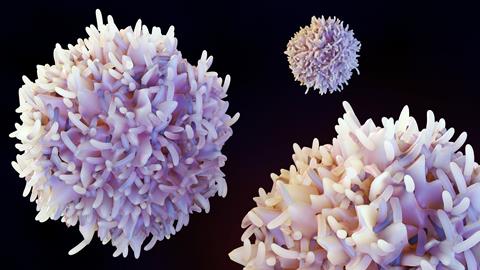
The new study, led by the University of Geneva and Lausanne University Hospital has, for the first time, captured the three-dimensional organisation of cytotoxic T lymphocytes – the body’s specialised ’killer’ cells – in a near-native state. The findings present new information that explains precisely how these cells function.
Cytotoxic T lymphocytes play a key role during infection or cancer. They attach themselves to harmful cells and form a tightly controlled exchange zone known as the immune synapse. Within this interface, they release toxic molecules that trigger the destruction of the target cell while sparing surrounding healthy tissue.
Cytotoxic T lymphocytes play a key role during infection or cancer
Although widely studied, the fine structure of this process at nanometre scale has been difficult to observe in intact human cells. Traditional imaging methods often force compromises between resolution, the size of the observable area and preservation of delicate cellular structures. Sample preparation itself has been challenging, as it can distort fragile biological material.
A technique to see the invisible
To overcome these limitations, the research team employed cryo-expansion microscopy, an imaging approach supported by the ISREC Foundation TANDEM programme.
“This technique involves instantaneously freezing cells at very high speed, placing them in a so-called vitreous state, where water solidifies without forming crystals and thus faithfully preserves biological structures,” said Virginie Hamel, Senior Lecturer in the Department of Molecular and Cellular Biology at the Faculty of Science of UNIGE. ”The samples are then physically expanded using an absorbent hydrogel, making it possible to observe their internal organisation with great precision while maintaining their near-native architecture.”
The method enabled scientists to examine immune cells with unprecedented clarity. At the contact point between the T cell and its target, they observed that the cell membrane forms a dome-like structure, shaped by adhesion interactions and the internal organisation of the cell.
The team also visualised cytotoxic granules – the components responsible for destroying target cells – in great depth. These structures were found to vary, sometimes containing one or more ’cores’ that concentrate the molecules needed to kill the target cell.
From cells to patients
Importantly, the researchers extended their approach beyond laboratory samples to human tumour tissues, offering a direct view of immune responses in a clinical setting.
“We extended this approach to human tumour tissues, making it possible to directly observe T lymphocytes infiltrating tumours and their cytotoxic machinery at the nanometer scale,” said Benita Wolf, Chief Resident and Associate Researcher in the Department of Clinical Oncology at CHUV, who co-led the study. ”This allows us to study immune responses directly in their clinical context and to better understand the mechanisms that determine their effectiveness.”
Importantly, the researchers extended their approach beyond laboratory samples to human tumour tissues
By providing a detailed 3D perspective, the research establishes a new reference framework for studying immune cell behaviour. Scientists believe this could help refine therapeutic strategies, particularly in immuno-oncology, by improving understanding of why immune responses succeed or fail in different patients.


No comments yet