


A new study has revealed that age-related clonal haematopoiesis accelerates aortic aneurysm growth through RANK/RANKL signalling, meaning that repurposing osteoporosis drugs targeting this pathway could provide the first pharmacological treatment option for the condition.
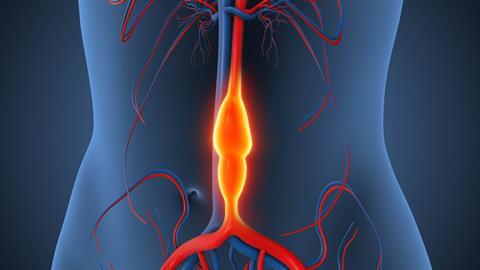
Aortic aneurysms, a dangerous condition that is characterised by abnormal enlargement of the aorta, are a major clinical challenge due to the lack of effective drug treatments. When they rupture, it often leads to sudden death and surgery is currently the only definitive option.
Now, a new study from researchers at Nagoya University has identified a potential new therapeutic pathway, suggesting that drugs already used to treat osteoporosis could slow or even stop the disease from progressing.
Clonal haematopoiesis linked to aneurysm progression
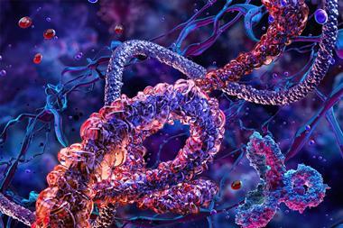
The team focused on clonal haematopoiesis, an age-related process in which blood-forming stem cells acquire genetic mutations. While this has been linked to conditions such as cardiovascular disease and osteoporosis, its role in aortic aneurysms was not defined.
The team focused on clonal haematopoiesis, an age-related process in which blood-forming stem cells acquire genetic mutations
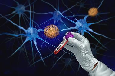
To investigate, the researchers analysed data from 44 patients scheduled for surgery for abdominal aortic aneurysms. Genetic testing and retrospective clinical data revealed that around 60 percent of patients had clonal haematopoiesis.
Importantly, these patients experienced significantly faster aneurysm growth compared with those without the condition. The findings suggest that clonal haematopoiesis, which can be detected through routine blood tests, may serve as a useful biological marker alongside existing imaging-based assessments.
Insights from animal models
To better understand the underlying mechanisms, the team turned to mouse models carrying Tet2 mutations, which drive clonal haematopoiesis. These mice showed more rapid aneurysm progression and greater enlargement of the aorta than controls.
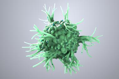
Detailed tissue analysis revealed damage to the aortic wall, including thinning and fragmentation of elastin fibres, along with increased infiltration of macrophages and degeneration of vascular smooth muscle cells.
Further research showed that these mutated macrophages exhibited features similar to osteoclasts, the cells responsible for breaking down bone. They also showed increased expression of molecules linked to tissue degradation, suggesting a mechanism by which they may accelerate aneurysm progression.
The researchers identified the RANK/RANKL signalling pathway as a key driver of this process. This pathway is already known to play a central role in osteoporosis. Notably, disabling the RANK gene in macrophages reduced abnormal cell behaviour and limited aortic enlargement in the animal models.
Repurposing existing drugs
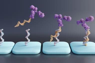
Building on these findings, the researchers tested whether drugs targeting this pathway could influence disease progression. Mice were treated with osteoporosis therapies, including anti-RANKL antibodies and the bisphosphonate alendronate.
The treatment significantly slowed aneurysm growth, raising the possibility of a non-surgical approach for patients.
Building on these findings, the researchers tested whether drugs targeting this pathway could influence disease progression
“These drugs could potentially be repurposed for clinical use as they are already FDA-approved and have established safety profiles,” said Jun Yonekawa, the study’s first author. “Our findings provide a rationale for exploring drug-based therapeutic strategies for aortic aneurysms.”
“Our hypothesis that vascular diseases may result from blood ageing enabled us to identify a mechanism underlying aortic aneurysms,” added Yoshimitsu Yura, the study’s corresponding author. “We hope these results will improve the prediction of the disease and support the development of treatments to halt progression.”
Towards better prediction and treatment
At present, clinicians rely on imaging features such as aneurysm size and growth rate to guide decisions about surgery. However, predicting which patients will experience rapid progression remains difficult.
This study highlights the potential of combining biological markers with imaging to improve risk assessment, while also potentially leading to drug-based therapies that could reduce the need for invasive procedures and lower mortality rates.


No comments yet