


Vitronectin has been identified as a critical mediator of macrophage reprogramming in idiopathic pulmonary fibrosis, revealing a previously unknown fibrotic mechanism uncovered through three-dimensional tissue culture modelling and validated in both animal models and patient samples.
Scientists have identified a protein that appears to trigger the scarring process behind idiopathic pulmonary fibrosis (IPF), which could help to create new treatments for the life-limiting lung disease.
The study was led by researchers from the University of Technology Sydney (UTS) and Monash University. It found that the protein vitronectin plays a key role in driving the abnormal scarring that characterises pulmonary fibrosis, offering a promising new therapeutic target.
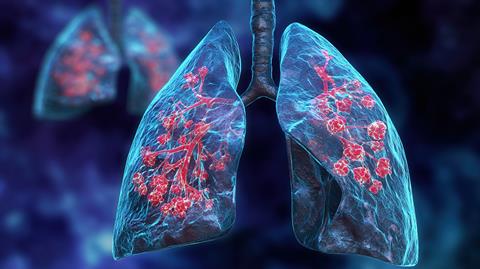
Pulmonary fibrosis causes scar tissue to build up in the lungs, making it increasingly difficult to breathe. Around 2,170 Australians are diagnosed each year with idiopathic pulmonary fibrosis, the most common form of the disease, which has no known cause and few treatment options.
Protein found to drive lung scarring
Researchers discovered that vitronectin can alter the behaviour of macrophages, immune cells that normally help repair damaged tissue after injury.
“In pulmonary fibrosis, the normal wound healing process in the body goes wrong. Instead of repairing damaged tissue, it starts to produce scar tissue in the lungs,” said Associate Professor Gang Liu from the UTS School of Life Sciences. “People with idiopathic pulmonary fibrosis have a very short survival time, usually only two to five years from diagnosis. Only two drugs are approved to treat it and neither of them can reverse the scarring and cure the disease.”
In pulmonary fibrosis, the normal wound healing process in the body goes wrong
Associate Professor Liu, co-first author of the study, said the team found that macrophages can become reprogrammed, causing them to promote fibrosis rather than healthy tissue repair.
“There’s an important type of immune cell – known as a macrophage – that helps repair tissue after an injury,” he said. “We discovered that these macrophages sometimes get reprogrammed to produce scarring rather than normal wound healing.”
3D model reveals new disease mechanism
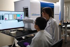
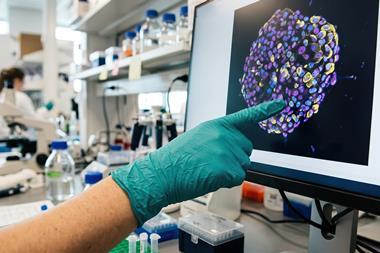
Senior author Associate Professor Katrina Binger from the Department of Biochemistry and Molecular Biology at Monash University developed a three-dimensional tissue culture system that recreated the fibrotic environment, allowing researchers to observe how vitronectin influences macrophage behaviour.
“We normally think of vitronectin as a structural protein that maintains the integrity of organs like the lungs. But we found it also can also act as a signal,” she said. “Vitronectin changes how macrophages produce energy and this drives them to have a heightened fibrotic state. This is a completely new mechanism to understand how fibrosis happens that was only possible by studying these cells in more natural, 3D environments.”
The findings from the laboratory model were also supported by animal studies and tissue samples from people with IPF.
Potential for future therapies
The researchers say the discovery provides a new direction for developing treatments that target the vitronectin-macrophage pathway, with the aim of slowing or preventing lung scarring.
“Understanding this mechanism is critical to identifying new therapeutic agents for fibrosis patients,” Associate Professor Liu said. “Now we can work to identify new drugs that can most effectively inhibit vitronectin so we can translate this research into clinical practice and help find a new cure for this debilitating disease.”
The researchers say the discovery provides a new direction for developing treatments that target the vitronectin-macrophage pathway
While further research is needed before new therapies become available, the findings are promising for people living with idiopathic pulmonary fibrosis, which has very few treatment options.








No comments yet