


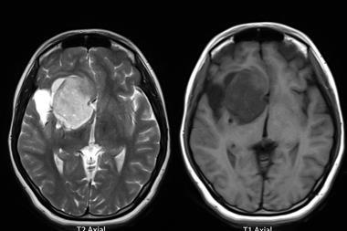
A cerebral organoid model has allowed scientists to see how Ebola virus persists and replicates in brain-like tissue for extended periods, infecting neurons, astrocytes and microglia while evading immune clearance.
Ebola virus can remain in the human body for months or even years after initial infection, hiding in immune-privileged regions such as the central nervous system where immune activity is limited. This ability to remain undetected raises concerns about viral relapse and, in rare cases, the possibility of renewed transmission.
A new study from the Icahn School of Medicine at Mount Sinai and the Bernhard Nocht Institute for Tropical Medicine (BNITM), in collaboration with international partners, has uncovered new information on how the virus survives in brain-like tissue.
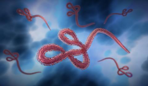
Ebola virus disease is a severe and often fatal illness caused by a filovirus. While some patients recover from the acute phase, the virus can remain in the body for extended periods. It has previously been detected in semen months or even a year after infection, and evidence suggests it can also persist in organs such as the brain.
Immune-privileged areas like the central nervous system are sites where immune responses are naturally reduced to protect delicate tissue. However, this reduced surveillance can also allow pathogens to persist, increasing the risk of later inflammatory complications and, in rare cases, transmission to others.
Brain organoid model offers new insights into viral persistence
Despite growing awareness of long-term Ebola virus persistence, the mechanisms that allow the virus to survive in the host are poorly understood. Researchers have questioned whether the virus remains within tissues or individual cells and whether it changes genetically to avoid immune detection.
To address these questions, the research team used a cerebral organoid model designed to mimic aspects of the human brain. The organoids were developed from human induced pluripotent stem cells which were guided to form three-dimensional structures containing multiple cell types found in the central nervous system.
Despite growing awareness of long-term Ebola virus persistence, the mechanisms that allow the virus to survive in the host are poorly understood
“These cerebral organoids enable us to investigate in detail the mechanisms that Ebola virus and other filoviruses use to persist in the human central nervous system. Through experiments in this model system, we can gain insights that help us improve our understanding of the long-term effects of persistence, like the severe and sometimes fatal inflammation seen in Ebola virus disease survivors with meningoencephalitis,” explains Dr Lina Widerspick, first author of the publication and former researcher at the BNITM.
Virus shows long-term replication in brain-like tissue
The researchers found that Ebola virus and related filoviruses including Sudan, Reston and Marburg viruses were able to replicate in cerebral organoids for up to 120 days.
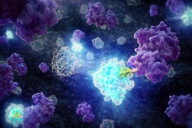
The virus infected multiple cell types including neurons and astrocytes. Microglia, the brain’s immune cells, were also drawn to infection sites and became infected. The virus spread both through direct cell-to-cell contact and by budding from infected cells, a standard mechanism of viral transmission.
This behaviour suggests a form of ’productive persistence’ in which the virus remains active and infectious rather than dormant.
Although infected organoids produced pro-inflammatory cytokines, the immune response was not sufficient to eliminate the virus. Over time, inflammation increased in late-stage cultures.
“We observed elevated immune and inflammatory responses in the late stages of cerebral organoid culture. We therefore conclude that a persistent Ebola virus infection in immune-privileged tissues can lead to local inflammation,” said Dr César Muñoz-Fontela, Head of the Virus Immunology Research Group at BNITM and co-last author of the study. ”This observation is consistent with the fact that some Ebola virus disease survivors develop inflammation of the eye, meninges or brain months after infection with Ebola virus.”
Genetic changes may help virus adapt to long-term survival
The team also identified defective viral genomes, mutations and viral particles that appeared during prolonged infection in organoids. These genetic changes are thought to influence replication and persistence.
“Many of these mutations had been proposed to reduce or prevent viral replication in naturally occurring infections. Because Ebola virus behaves similarly in this model system to how it does in human infections, this underscores the suitability of our cerebral organoids for investigating filovirus persistence,” explains Dr Gustavo Palacios, Professor of Microbiology at the Icahn School of Medicine, co-last author of the publication and an expert on Ebola virus genomics.
Many of these mutations had been proposed to reduce or prevent viral replication in naturally occurring infections
Some mutations observed had not previously been reported in Ebola virus disease survivors, suggesting further research is needed to understand their role in persistence.
“Our work in human cerebral organoids highlights the potential of this model system to investigate persistent infections in immune-privileged tissues,” said Dr Palacios. “Further studies are now important to investigate the long-term interactions between virus and host, expanding our studies towards less-studied filoviruses like Reston, Taï Forest, Bombali and Bundibugyo virus and to deepen our understanding of filoviral persistence mechanisms.”


No comments yet