


How does Ebola virus survive long after recovery? A new study using human cerebral organoids explores viral persistence in neural tissue and the growing role of organoid models in drug discovery research.

One of the most challenging aspects of Ebola virus is how the virus survives long after acute infection has resolved. Infectious Ebola virus has been detected months or even years after recovery, indicating that viral reservoirs can remain within the body despite an apparently successful immune response.
Particular attention has focused on the central nervous system. The brain is considered an immune-privileged organ, where inflammatory responses are tightly controlled to minimise damage to sensitive neural tissue. While this protection is essential for normal function, it can complicate efforts to understand how viruses are cleared from the tissue and whether infectious virus can remain behind after recovery.
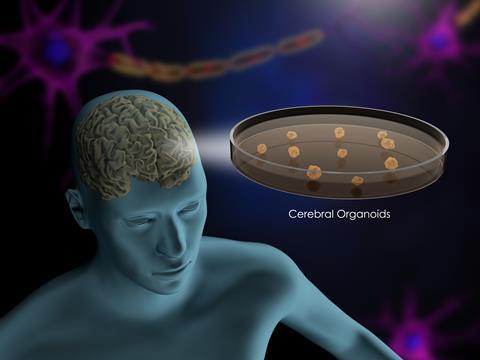
A new study led by researchers from the Icahn School of Medicine at Mount Sinai and the Bernhard Nocht Institute for Tropical Medicine used cerebral organoids derived from human induced pluripotent stem cells (iPSCs) to investigate how Ebola virus persists in the central nervous system.
Using this model, the team showed that Ebola virus can establish long-term infection in human neural tissue while continuing to replicate, spread and accumulate genetic changes.
Why Ebola persistence remains a challenge
Studying viral persistence in the human brain presents significant challenges. Access to patient tissue is extremely limited, while animal models cannot fully reproduce the complexity of human neurological disease. As a result, researchers still do not fully understand which cell types support long-term infection, whether the virus continues to replicate during persistence, how local immune responses influence disease progression, or which viral genetic changes emerge over time.
Addressing these questions requires experimental systems capable of supporting long-term infection while capturing key features of human tissue biology. This is where cerebral organoids have begun to provide researchers with a valuable new tool.
Building a model of the human brain
To investigate these mechanisms, the research team used cerebral organoids generated from iPSCs.
The researchers directed iPSCs to form spherical brain-like structures composed of several central nervous system cell types, including neurons, astrocytes and other supporting cells found within human neural tissue.
Although cerebral organoids cannot replicate the full complexity of the human brain, they offer a more physiologically relevant system for investigating long-term infection than traditional cell culture models. This makes them particularly useful for studying biological processes that are difficult to observe directly in patients, including viral persistence within the central nervous system.
Productive persistence rather than viral dormancy
One of the most significant findings from the study was that Ebola virus remained actively infectious throughout the persistence phase.
Researchers demonstrated that Ebola virus, together with related filoviruses including Sudan virus, Reston virus and Marburg virus, could replicate within cerebral organoids for up to 120 days.
Importantly, the virus did not simply remain dormant within infected cells. Instead, investigators observed continued viral replication and spread throughout the tissue.
Two transmission mechanisms were identified. The virus spread directly between neighbouring cells and also used conventional viral budding, whereby newly formed viral particles are released from infected cells and infect surrounding targets.
The study showed that Ebola virus does not simply remain present within neural tissue. Instead, it continues to replicate and spread during persistence, indicating that viral reservoirs within the central nervous system may remain active long after acute infection has resolved.
Multiple brain cell populations support infection
The study also showed that Ebola virus is capable of infecting multiple neural cell types. Researchers detected infection in neurons and astrocytes, both essential to central nervous system function, while microglia – the brain’s resident immune cells – were recruited to infected regions and also became infected.
The ability to infect several cellular compartments may help explain the neurological complications reported in some Ebola survivors. It also highlights a challenge for therapeutic development. Viral reservoirs distributed across different cell populations may be more difficult to eliminate than infections confined to a single cellular niche, particularly if drug penetration or antiviral activity varies between cell types.
Implications for antiviral development
Although the study did not evaluate antiviral candidates, its implications extend beyond understanding Ebola virus biology.
One of the persistent challenges in infectious disease research is the lack of human-relevant systems for studying long-term infection. Viral persistence can be difficult to model in patients and is often only partially captured in animal studies, limiting researchers’ ability to investigate how pathogens survive within specific tissues over time.
By sustaining productive Ebola virus infection, the cerebral organoids provided a platform for examining viral replication, host responses and genetic changes within human neural tissue. The authors suggest that such models could support the reassessment of antiviral strategies by enabling researchers to study persistent infection in a controlled human-derived environment.
What challenges remain?
As with any organoid model, there are limitations. Cerebral organoids lack vascularisation, systemic immune responses and the full cellular diversity of the human brain, while several mutations identified during infection have yet to be observed in Ebola survivors and require further investigation.
Even so, maintaining productive Ebola virus infection for up to 120 days allowed the researchers to examine viral replication, host responses and genetic changes within human neural tissue over an extended period. For a field where direct access to infected brain tissue is exceptionally limited, that represents a valuable experimental opportunity. The study illustrates how stem cell-derived models can be used to investigate viral reservoirs in human tissue and may support future efforts to evaluate therapies designed to eliminate residual virus following infection.


No comments yet