

Many antibody therapies fail to reach all cancer cells within solid tumours. Researchers have developed a spatial biology technique that maps antibody distribution alongside the tumour microenvironment to investigate why.
Antibody therapies have become an important part of cancer treatment, recognising proteins expressed on tumour cells to selectively target cancerous tissue. This has led to the development of several major therapeutic classes, including immune checkpoint inhibitors, bispecific antibodies and antibody-drug conjugates (ADCs). However, many solid tumours remain difficult to treat. Response rates are often modest and expression of the intended molecular target alone does not guarantee a meaningful clinical response.
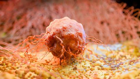
One explanation is that therapeutic antibodies do not always reach every cancer cell within a tumour. Solid tumours are highly complex tissues comprising cancer cells, blood vessels, immune cells, fibroblasts and extracellular matrix proteins. Together, these components form the tumour microenvironment, which can influence how effectively drugs penetrate different regions of the tumour.
Until recently, scientists have lacked tools capable of showing exactly where therapeutic antibodies travel once they enter a tumour and how the surrounding tissue influences their activity.
A new study published in Nature Biotechnology describes a technique called single-cell spatial pharmacobiology (SSP) that aims to address this challenge. By combining fluorescent imaging with multiplex spatial proteomics, the researchers examined where therapeutic antibodies localise within human tumours, which cells they bind to and how tumour architecture influences drug distribution.
Why drug delivery remains a challenge
Developing an effective antibody therapy involves much more than identifying the right molecular target. Once administered, the drug must leave the bloodstream, enter the tumour, diffuse through dense tissue and bind to its target in sufficient quantities to produce a biological effect.
Traditional methods can confirm that an antibody reaches the general location of a tumour. Blood sampling provides information about systemic drug exposure, while imaging techniques such as positron emission tomography (PET) can show uptake across an entire tumour. However, these approaches lack the spatial resolution needed to determine whether the drug reaches individual cancer cells or becomes trapped within surrounding tissue.
Tumours are rarely uniform. Cells at the tumour edge may receive abundant oxygen, nutrients and blood supply, while cells deeper within the tumour often exist in hypoxic, densely fibrotic environments. These differences can create regions where therapeutic antibodies penetrate effectively alongside neighbouring areas that receive very little drug. Understanding these spatial differences has become an important objective in cancer drug development.
Bringing spatial biology into pharmacology
Over the past decade, spatial biology has emerged as one of the fastest-growing areas of biomedical research. Unlike conventional sequencing or bulk tissue analysis, spatial technologies preserve the physical location of cells within intact tissue. Researchers can therefore examine not only which genes or proteins are present, but also where they are expressed and how neighbouring cells interact.
Researchers at Stanford University developed SSP, an approach designed to study the behaviour of therapeutic drugs directly within human tumours. Rather than analysing tissue after treatment using indirect measurements, SSP combines high-resolution imaging of a fluorescently labelled therapeutic antibody with multiplex spatial proteomics to examine drug distribution alongside the tumour microenvironment.
For this proof-of-concept study, the researchers investigated panitumumab, a monoclonal antibody that targets epidermal growth factor receptor (EGFR), a protein commonly overexpressed on the surface of cancer cells. The antibody was labelled with a near-infrared fluorescent dye before being administered to patients participating in phase I clinical trials.
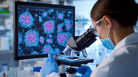
After surgical tumour removal, the tissue was analysed using CODEX, a multiplex imaging platform capable of simultaneously detecting dozens of proteins within the same tissue section. Computational image registration then aligned the fluorescent drug signal with the spatial protein maps at single-cell resolution. The resulting images showed where therapeutic antibodies travelled within intact human tumours and how their localisation related to blood vessels, immune cells, fibroblasts and extracellular matrix proteins.
Antibody delivery varies dramatically within tumours
Applying SSP to head and neck squamous cell carcinoma (HNSCC) and pancreatic ductal adenocarcinoma (PDAC) revealed extensive variability in antibody distribution. Differences were observed not only between patients, but also between separate regions of the same tumour.
The researchers found that many tumour cells expressing EGFR were not reached by the therapeutic antibody. In HNSCC, only a minority of tumour cells both expressed EGFR and showed detectable antibody binding, while many EGFR-positive cells exhibited little or no evidence of drug engagement. Rather than being evenly distributed throughout the tumour, antibody-bound cells were concentrated near blood vessels and around the tumour periphery. Cells located deeper within tumour nests were much less likely to be reached.
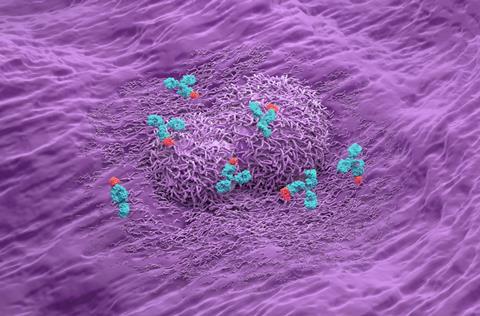
The findings indicate that target expression alone may not accurately predict treatment response. Even if a tumour contains abundant levels of the intended target, the therapy may fail if the antibody cannot physically access those cells.
Bulk measurements of drug concentration might suggest that a therapy has successfully reached a tumour, while single-cell spatial analysis reveals that substantial regions remain effectively untreated.
The tumour microenvironment acts as a physical barrier
To understand why some tumour regions were inaccessible, the researchers turned their attention to the tumour microenvironment.
Solid tumours are surrounded by an extracellular matrix (ECM), a network of structural proteins that provides physical support for tissues. Although the ECM is essential for normal tissue function, cancers often remodel this matrix, producing dense fibrotic regions that may limit drug movement.
Among the ECM proteins examined, periostin emerged as the strongest correlate of poor antibody penetration. Regions enriched in periostin consistently showed reduced levels of therapeutic antibody, suggesting that dense periostin-rich matrix structures may act as physical barriers restricting diffusion through the tumour.
The researchers also identified fibroblast activation protein (FAP)-positive cancer-associated fibroblasts (CAFs) as another feature associated with reduced antibody penetration. These specialised stromal cells produce many extracellular matrix components and were frequently found adjacent to periostin-rich regions. Together, FAP-positive fibroblasts and periostin-rich matrix formed conserved stromal niches associated with reduced antibody penetration in both HNSCC and PDAC.
The fact that similar spatial patterns were observed across two biologically distinct tumour types suggests these stromal barriers may represent common mechanisms limiting therapeutic antibody delivery in solid cancers. The PDAC comparison was based on four patients matched for antibody dose. The authors therefore present these findings as supporting evidence for a shared mechanism rather than definitive proof.
Implications for antibody drug discovery
The ability to visualise therapeutic antibody distribution at single-cell resolution could support several stages of drug discovery.
During preclinical optimisation, researchers often focus on improving binding affinity, stability or pharmacokinetic properties. However, these findings indicate that successful therapies must also overcome physical barriers within the tumour microenvironment. Spatial pharmacology could therefore be incorporated into antibody candidate selection.
The approach may also prove valuable for ADCs, where insufficient delivery could prevent the cytotoxic payload from reaching its intended target. Similarly, immune checkpoint inhibitors may fail not because immune cells cannot respond, but because the therapeutic antibody never adequately penetrates the relevant tumour regions.
By identifying stromal features associated with poor drug delivery, SSP may help researchers distinguish between biological resistance and inadequate tumour exposure.
Supporting biomarker discovery and precision oncology
SSP may also support biomarker discovery. Many predictive biomarkers currently focus on molecular target expression alone. However, the study demonstrates that target abundance may not always correspond to therapeutic exposure. Incorporating spatial information could therefore improve patient stratification by identifying tumours whose architecture is more likely to permit effective antibody penetration.
The authors also suggest that spatial pharmacobiology could support early clinical trials by directly measuring drug delivery and target engagement in human tissue. Rather than relying solely on conventional pharmacokinetic measurements, investigators could evaluate how candidate therapies behave within individual tumours, potentially providing earlier evidence of therapeutic activity or resistance mechanisms.
What comes next?
Although this study focused on an EGFR-targeting antibody, the underlying framework is broadly applicable. The researchers propose that SSP could be adapted to investigate immune checkpoint inhibitors, antibody-drug conjugates and other antibody-based therapeutics.
Further studies will be needed to determine whether these findings extend to additional tumour types and antibody therapies. However, the study demonstrates how spatial biology and pharmacology can be combined to investigate therapeutic antibody distribution and the influence of tumour architecture on drug penetration.







No comments yet