


FcRn plays a central role in regulating the half-life of IgG antibodies and albumin, making it a critical target in both antibody engineering and autoimmune disease therapy. This article explores the biology of FcRn, the use of HMEC-1 cells for measuring FcRn-mediated IgG recycling, and the assays used to support the development of next-generation biologics and FcRn-targeted therapeutics.
1/ What is the biological function of FcRn?
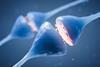
Neonatal Fc receptor for IgG (FcRn) is a heterodimeric protein similar in structure to MHC class I that binds to the Fc region of immunoglobulin G (IgG). It consists of the FcRn heavy chain (encoded by FCGRT) associated with β2-microglobulin (B2M) and is expressed in over 25 tissue types, with high expression levels observed in the spleen and intestine.
FcRn contributes to effective humoral immunity by protecting IgG antibodies from degradation, recycling them and extending their half-life in circulation. It also regulates the homeostasis of serum albumin.
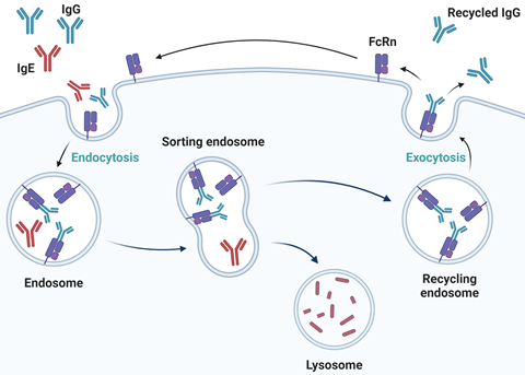
FcRn plays a vital role in regulating the level of albumin and IgG antibodies in circulation by binding albumin and the Fc region of an IgG at low pH (about pH 6.0) in endosomes. It then diverts them from lysosomal degradation and recycles them for release into the neutral pH (pH 7.0 to 7.4) of the extracellular compartment.
2/ How is FcRn function exploited for antibody engineering?
The IgG antibody stabilisation function of FcRn can be exploited when engineering therapeutic antibodies: mutations introduced into the Fc fragment can increase binding to FcRn, which improves their half-life and therefore their therapeutic efficacy.
For example, an antibody cocktail that contains Fc mutations – and thus an extended half-life (Evusheld) – has been used to treat COVID-19. First-in-class drug Enbrel (etanercept) contains the extracellular domain of TNF receptor 2 fused to a human IgG1 Fc domain, which contributes to its prolonged serum half-life.
Measuring FcRn-dependent IgG antibody recycling is therefore a key step in developing and characterising novel therapeutic antibodies, Fc-engineered biologics and biosimilars. Researchers often use HMEC-1 cells for this purpose.
3/ Why are HMEC-1 cells used to perform IgG antibody recycling assays?
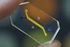
The HMEC-1 cell line (human microvascular endothelial cells, immortalised) is one of the most widely used in vitro models for evaluating the internalisation and recycling of therapeutic IgG antibodies. As an endothelial cell system, it is highly physiologically relevant because endothelial cells are considered one of the major sites of FcRn-mediated IgG recycling in vivo. HMEC-1 cells express the neonatal Fc receptor (FcRn), enabling the study of key processes involved in IgG trafficking, including cellular uptake, endosomal sorting and recycling back to the cell surface. This makes them well suited for assessing the recycling efficiency of therapeutic antibodies and engineered Fc variants.
In addition to their biological relevance, HMEC-1 cells provide several practical advantages. As an immortalised cell line, they offer a stable and reproducible experimental system with consistent FcRn expression and assay performance over time. In contrast, primary endothelial cells can be difficult to culture and often exhibit donor-to-donor variability. HMEC-1 cells also grow as an adherent monolayer, simplifying washing and media exchange steps, while their robust uptake and recycling kinetics generate strong signal-to-noise ratios.
Together, these characteristics make HMEC-1 cells a valuable platform for comparing antibody candidates, screening Fc-engineered variants and studying FcRn-mediated recycling mechanisms.
4/ How does the FcRn: IgG Recycling HMEC-1 Cell Pool work?
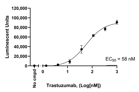
BPS Bioscience’s cell-based IgG antibody recycling assays are designed to evaluate FcRn-mediated antibody trafficking and recycling activity in physiologically relevant HMEC-1 cells. Sometimes termed HERA (human endothelial cell-based recycling assay), these assays support lead optimisation and functional characterisation workflows by providing quantitative data on IgG binding to FcRn and recycling behaviour.
The assays use HMEC-1 cells genetically engineered to express FcRn, for robust, stable expression of FCGRT and B2M. The cells were extensively validated by measuring the recycling of well-known IgG1 antibodies such as nivolumab, rituximab, trastuzumab, basiliximab and alirocumab.
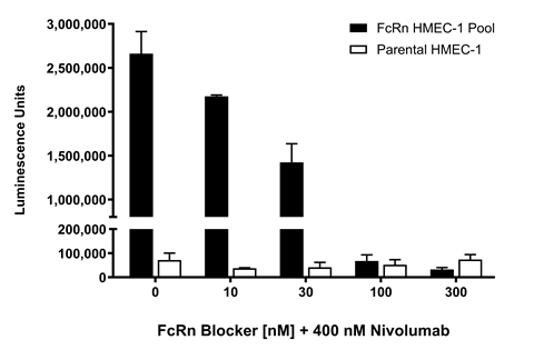
5/ When is it desirable to block IgG binding to FcRn?
FcRn itself is a therapeutic target for some autoimmune diseases since disrupting the FcRn/IgG interaction is expected to increase IgG antibody clearance, including clearance of autoantibodies, thereby decreasing their concentration in patients. The first FDA-approved drug targeting FcRn (efgartigimod) is an Fc fragment decoy that provided proof-of-concept and is now used to treat the autoimmune disease myasthenia gravis.
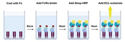
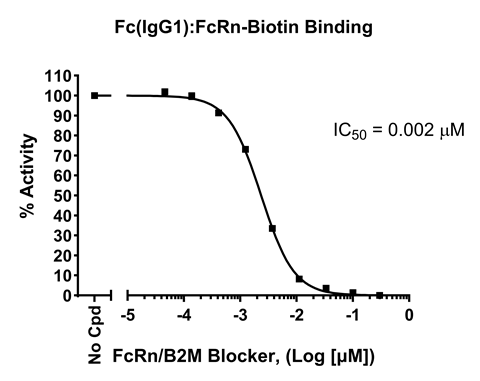
To facilitate the screening and optimisation of such FcRn blockers, BPS Bioscience has designed colorimetric (#78501) and chemiluminescent (#82652) ELISA kits that directly measure the binding of FcRn to Fc-containing biologics.


No comments yet