

Electron microscopy details infection mechanisms of coronaviruses
Posted: 29 February 2016 | Victoria White | No comments yet
High-resolution cryo-electron microscopy and supercomputing have now made it possible to analyse in detail the infection mechanisms of coronaviruses…
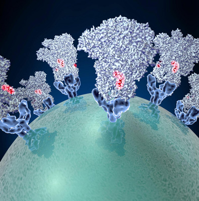
Coronaviruses employ molecular tactics to infect cells. CREDIT: Veesler Lab/University of Washington
High-resolution cryo-electron microscopy and supercomputing have now made it possible to analyse in detail the infection mechanisms of coronaviruses.
A research team that included scientists from the University of Washington (UW), the Pasteur Institute and the University of Utrecht has obtained an atomic model of a coronavirus spike protein that promotes entry into cells. Analysis of the model is providing ideas for specific vaccine strategies.
These viruses, with their crowns of spikes, are responsible for almost a third of mild, cold-like symptoms and atypical pneumonia worldwide, David Veesler, UW assistant professor of biochemistry, explained. But deadly forms of coronaviruses emerged in the form of SARS-CoV (severe acute respiratory syndrome coronavirus) in 2002 and of MERS-CoV (Middle East respiratory syndrome coronavirus) in 2012 with fatality rates between 10% and 37%.
These outbreaks of deadly pneumonia showed that coronaviruses can transmit from various animals to people. Currently, only six coronaviruses are known to infect people, but many coronaviruses naturally infect animals. The recent deadly outbreaks resulted from coronaviruses overcoming the species barrier. This suggests that other new, emerging coronavirus with pandemic potential are likely to emerge.
The ability of coronaviruses to attach to and enter specific cells is mediated by a transmembrane spike glycoprotein. It forms trimers decorating the virus surface. The structure the researchers studied is in charge of binding to and fusing with the membrane of a living cell. The spike determines what kinds of animals and what types of cells in their bodies each coronavirus can infect.
Using state of the art, single particle cryo-electron microscopy and supercomputing analysis, Veesler and his colleagues revealed the architecture of a mouse coronavirus spike glycoprotein trimer. They uncovered an unprecedented level of detail. The resolution is 4 angstroms, a unit of measurement that expresses the size of atoms and the distances between them and that is equivalent to one-tenth of a nanometre.
“The structure is maintained in its pre-fusion state, and then undergoes major rearrangements to trigger fusion of the viral and host membranes and initiate infection,” Veesler explained.
Accessibility of peripheral peptide may influence vaccine design
The coronavirus fusion machinery is reminiscent of the fusion proteins found in another family of viruses, the paramyxoviruses, which include respiratory syncytial virus (RSV) as well as the viruses that cause measles and mumps. This resemblance implies that the coronavirus and paramyxovirus fusion proteins could employ similar mechanisms to promote viral entry and share a common evolutionary origin.
The researchers also compared crystal structures of parts of the spike protein in mouse and human coronaviruses. Their findings provide clues as to how the molecular structure of these protein domains might influence which specific animal species the virus is able to infect.
The researchers also analysed the structure for possible targets for vaccine design and anti-viral therapies. They observed that the outer edge of the coronavirus spike trimer has a fusion peptide that is involved in viral entry into host cells. The easy accessibility of this peptide, and its expected similarity among a number of coronaviruses, suggests possible vaccine strategies to neutralise a variety of these viruses.
There may be a way, the researchers noted, to elicit broadly neutralising antibodies recognising this peripheral peptide. The physical structure of the fusion peptide inspires ideas for the design of proteins that would disable it.
Veesler explained: “Small molecules or protein scaffolds might eventually be designed to bind to this site to hinder insertion of the fusion peptide into the host cell membrane and to prevent it from undergoing changes conducive to fusion with the host cell. We hope that this might be the case, but much more work needs to be done to see if it is possible.”
Related conditions
Middle East respiratory syndrome coronavirus (MERS-CoV)
Related organisations
Institut Pasteur, University of Utrecht, Washington University