


Two cancer drugs appeared almost identical in conventional screening, but a new 3D Cell Painting method exposed striking differences in their effects, raising questions about what 2D models might miss.
Researchers at Uppsala University in Sweden have developed a 3D version of Cell Painting that can identify differences in how cancer cells respond to drugs that appear almost identical in conventional 2D cultures.
The study, published in Nature Communications, demonstrates how combining 3D cell models with high-content imaging and single-cell analysis could improve the assessment of drug activity during early discovery.
Further analysis revealed differences in how the drugs affected DNA damage, cell division and the p53 stress response, demonstrating the additional information that 3D screening can provide during early drug discovery.
Adapting Cell Painting for 3D drug screening
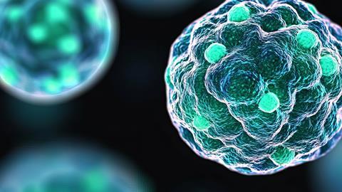
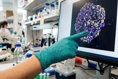
Cell Painting is an imaging technique used in drug discovery to measure changes in cellular structures following treatment with compounds. Fluorescent dyes highlight components such as the nucleus, mitochondria and cytoskeleton, allowing researchers to identify changes in cell morphology.
Although widely used in high-content screening, the technique has traditionally relied on cells grown in two-dimensional layers, which do not fully reproduce the organisation of cells in tissues.
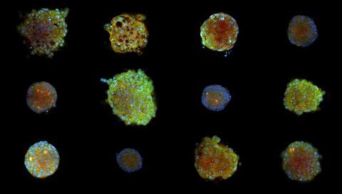
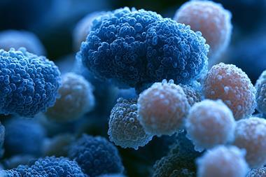
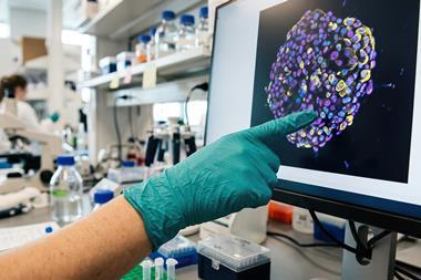
To address this limitation, the Uppsala team grew colorectal cancer cells as small three-dimensional spheroids. Using a clearing solution containing sugar and glycerol, they made the spheroids transparent enough to image individual cells throughout the structures.
The researchers then extracted thousands of morphological features from individual cells, allowing drug-induced changes to be analysed without losing the detail normally obscured in 3D samples.
“Cells behave differently when they grow in three dimensions, but most screening methods either use flat cultures or collapse a whole 3D sample into one picture. We wanted to keep the detail of single cells while still being able to test many drugs at once,” said Christa Ringers, joint first author of the study.
Revealing differences between cancer drugs
Among the 52 compounds tested, the researchers identified a notable difference between 5-fluorouracil, a chemotherapy drug, and olaparib, a PARP inhibitor.
Although the two drugs produced nearly indistinguishable morphological profiles in 2D cultures, their effects became clearly distinguishable in 3D spheroids when analysed at the single-cell level. These differences were not apparent when measurements were averaged across entire spheroids.
Further experiments found that 5-fluorouracil caused less DNA damage than olaparib but was more effective at preventing cell division. Gene expression analysis linked this response to stronger activation of the p53 pathway, which regulates cellular responses to stress and DNA damage.
The team also tested the method with collaborators at AstraZeneca to assess whether it could be reproduced outside the laboratory where it was developed.
The findings suggest that 3D Cell Painting could provide additional information about compound activity and mechanisms of action, particularly when drugs produce similar responses in conventional screening assays.
Although the research was conducted using two laboratory cancer cell lines and does not establish how the drugs behave in patients, the researchers hope to extend the technique to more complex models, including patient-derived organoids.
Reference
Ringers C, et al. High-content morphological profiling by Cell Painting in 3D spheroids. Nat Commun. 2026. doi: 10.1038/s41467-026-78596-5.







No comments yet