On-demand webinar

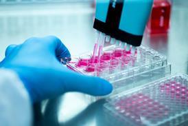
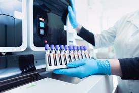
Complete aggregate and particle characterisation of protein, gene and cell therapies

Join us to learn about the importance of subvisible particle characterisation for better product stability, ensuring patient safety from early-phase development through USP 788 lot release testing.

Register to join this webinar discussion for free!

In this webinar, our key speaker Dikran Khachadourian, will discuss how Aura® incorporates the techniques of Backgrounded Membrane Imaging (BMI) and Fluorescence Membrane Microscopy (FMM) to allow for the complete characterisation and identification of subvisible particles in protein, gene, and cell therapy products requiring as little as 5 µL of sample. We’ll highlight applications ranging from developability assessment to product development and release.

Key learnings

  • Understand the importance of testing the purity and stability of drug products to ensure efficacy and patient safety.
  • Learn why Backgrounded Membrane Imaging (BMI) has become an emerging technique for lot release testing and generating USP 788-compliant data.
  • Master particle and product characterization as early as early-phase development using only 5uL of sample leveraging a modernised approach of membrane microscopy.
  • Explore how Fluorescence Membrane Microscopy (FMM) can be used to differentiate between subpopulations of particulates, such as protein aggregates, polysorbate degradants, and extrinsic contaminants.

Secure your place now! Complete your details in the box and join us on 13th November at 3.00pm.

CONTEXT

In all biological products, distinguishing aggregated API from other particle types matters for understanding the root cause of instability. Ideally, particle characterisation should begin in early-phase development to determine the best drug candidate through formulation development to ensure product stability and, finally, in lot release for product purity and safety. Current subvisible particle characterisation methods have been unreliable, slow, require significant sample volume, and are difficult to use across different therapeutics. More alarming is these antiquated methods often undercount biological aggregates and other contaminants, putting product stability and patient safety at significantly high risk. Membrane microscopy (a modernised form of automated light microscopy) is an alternative approach to this problem.

Being USP 788 and 1788 compatible, this method can be ultra-low volume while providing higher throughput to rapidly size, count, and characterise biological particles and identify them as proteins, non-proteins, polysorbate degradants, cellular aggregates, or other types of molecules.

SPEAKER

Dikran Khachadourian

Dikran Khachadourian , Field Application Scientist, Halo Labs 

Dikran Khachadourian is a Field Application Scientist at Halo Labs. He completed his undergraduate studies at UC San Diego, obtaining a bachelor’s degree in Biochemistry and Cell Biology. His interest in the biological sciences led him to pursue a master’s degree in biology. After spending time in academia as a graduate researcher and educator, Dikran joined the Halo Labs team to apply his background to the ongoing advancements in biotechnology. 


FAQs

FAQs

Is the webinar free? 

Yes – there is no charge to watch the webinar, either live or on-demand. 

When will the webinar take place? 

13th November 2023 at 3.00pm GMT. 

Can I watch it later? 

The webinar will become available to watch on-demand shortly after the live webinar takes place. 

What are the benefits of attending live? 

You’ll be able to ask the speakers your questions, which will be answered live in the Q&A towards the end of the session. 

How long will the webinar be? 

This webinar will last up to an hour. 

What do I need to watch this webinar? 

All you need is a computer with an internet connection. We recommend using headphones if possible if you’re in an office environment.

ABOUT US

Halo Labs is a venture-backed scientific instrumentation and consumable company that commercializes high-throughput biopharmaceutical formulation and quality control tools for particle analysis using state-of-the-art custom optics and image processing techniques.

Halo Labs knows particles. The Aura, our flagship product, can count and characterize subvisible particles – and tell you whether they are proteinaceous or non-proteinaceous. A simple, plate-based approach enables low-volume, high-throughput, fully automated particle imaging and analysis at any stage from developability assessment through quality control.

Halo Labs’ core mission has been to provide researchers with unprecedented insights into the particles present in their processes by leveraging our core scientific competency: optical imaging on membranes. Over the years, Halo Labs has been the recipient of many prestigious awards, and has received over $6M in research grants from agencies including DARPA, FDA, NSF, NIST and NIH. Today, we dedicate our collective experience and novel technology to empower formulation researchers to learn more about the stability and quality of their biological drugs faster and with more accuracy than ever before.

Our institutional partners include Research Corporation Technologies (RCT), BioAdvance, Broad Oak Capital Partners, and Ben Franklin.