


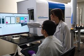
Researchers have used the GAGE-seq technique and an AI model called Hicformer to demonstrate that the three-dimensional folding of the genome is significantly disrupted in brain cells affected by Alzheimer’s disease – a finding that could help prioritise new targets for therapeutic investigation.
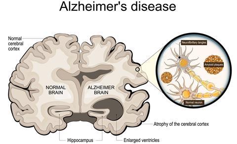
Researchers have uncovered a previously overlooked feature of Alzheimer’s disease by showing that the three-dimensional organisation of the genome is altered in brain cells affected by the condition, which could help guide the development of future treatments.
The international study was lead by researchers from Carnegie Mellon University’s School of Computer Science, the University of Pittsburgh School of Medicine and the University of Washington. By combining single-cell analysis, spatial transcriptomics and artificial intelligence, the team linked changes in genome architecture to altered gene activity and brain tissue organisation in Alzheimer’s disease.
Connecting genome structure to disease
Rather than examining genes in isolation, the researchers investigated how the genome folds inside cells and how this influences gene activity. They analysed postmortem tissue from the prefrontal cortex of people with and without Alzheimer’s disease who had participated in a long-term dementia study and donated their brains for research.
The team used GAGE-seq, a technique that measures gene expression and three-dimensional genome contacts within the same cell. These findings were then integrated with spatial transcriptomic maps of intact brain tissue, enabling the researchers to examine molecular changes within their tissue environment.
Rather than examining genes in isolation, the researchers investigated how the genome folds inside cells and how this influences gene activity
“Alzheimer’s disease cannot be understood one layer at a time,” said Jian Ma, the Ray and Stephanie Lane Professor of Computational Biology at Carnegie Mellon University, who led and supervised the study. ”The genome’s 3D structure is a fundamental regulatory layer that helps to connect DNA sequence to gene activity. By integrating genome folding, cell state and tissue context, we can move beyond cataloging disease-associated changes toward understanding how they fit together and which mechanisms to test next.”
AI model identifies altered genome organisation
A key element of the research was Hicformer, an artificial intelligence model developed to predict gene activity by combining DNA sequence data with information on genome folding and local three-dimensional contact maps.
Using this approach, the researchers found that the separation between active and inactive regions of the genome was less distinct in brain cells from people with Alzheimer’s disease, a phenomenon they describe as ’increased compartment mingling’. The affected cells also displayed fewer short-range genome contacts and more long-range interactions, changes that were associated with reduced overall gene activity.
The researchers found that the separation between active and inactive regions of the genome was less distinct in brain cells from people with Alzheimer’s disease
These structural alterations were linked to reduced neuronal and synaptic programmes, altered metabolic and stress responses and senescence-related programmes in microglia.
“Our study represents a major advance in understanding what goes wrong in Alzheimer’s disease,” said Hansruedi Mathys, Assistant Professor of Neurobiology at the University of Pittsburgh’s Department of Neurobiology, who directed the Pittsburgh arm of the study. ”We know the classic hallmarks of Alzheimer’s disease – accumulation of amyloid-beta plaques and tau tangles – but our results establish higher-order chromatin alterations as a component of the molecular pathology associated with the disease, which currently affects seven million Americans, a number that continues to grow.”
Studies such as this highlight how understanding disease often requires more than studying cells in isolation. Our latest report explores how spatial biology is revealing aspects of disease biology that cannot be captured through individual cells alone, and what that could mean for biomarker discovery, immunotherapy and drug development.
Providing a framework for future therapies
The researchers also found that changes in genome organisation corresponded with alterations in gene activity and the arrangement of brain cells across intact tissue, highlighting three-dimensional genome architecture as an important component of Alzheimer’s biology.
“Measuring gene activity and genome folding in the same cell allows us to directly connect chromosome structure with disease-related gene programs,” said Yang Zhang, a project scientist in Carnegie Mellon University’s Computational Biology Department and co-lead author of the study. ”Across several kinds of brain cells, this paired view revealed a consistent signature of 3D genome reorganisation in Alzheimer’s disease and helped us prioritise regulatory regions for future mechanistic and therapeutic investigation.”
The researchers say the findings provide a framework for future studies to determine whether changes in genome structure contribute directly to Alzheimer’s disease and whether they could become targets for new therapies.











No comments yet