

Scientists reveal how signals from pathogenic bacteria reach danger sensors of cells
Posted: 29 September 2016 | Niamh Louise Marriott, Digital Content Producer | No comments yet
Signals from infectious bacteria gain entry into the cytoplasm of host cells to activate disease-fighting inflammasomes, protein machines in the cell that serve as “danger sensors” for infection and which set in motion rapid immune defences against pathogens…
Researchers at St. Jude Children’s Research Hospital have discovered the way signals from infectious bacteria gain entry into the cytoplasm of host cells to activate disease-fighting inflammasomes, protein machines in the cell that serve as “danger sensors” for infection and which set in motion rapid immune defences against pathogens.
The researchers, led by Thirumala-Devi Kanneganti, PhD, a member of the St. Jude Department of Immunology, showed that the interferon-inducible protein called IRGB10 is essential for the activation of inflammasomes.
“Researchers have known for some time that bacterial ligands or signals must gain entry into the cytoplasm of cells to activate inflammasomes. However, the mechanism by which these signals are liberated and presented to disease-fighting inflammasome sensors has not been clear,” said Kanneganti.
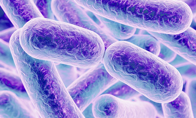
In the past 15 years, around five different types of pathogen-fighting inflammasomes have been identified in cells. In the current study, the scientists looked at how different bacteria triggered defence responses in bone marrow-derived macrophages in mice.
The scientists revealed that IRGB10 is essential for the activation of two inflammasomes. When cells were exposed to the pathogen Francisella novicida, the AIM2 inflammasome, a sensor that detects DNA from invading bacteria, was activated.
- novicida is a highly infectious pathogen that causes rabbit fever, a potentially fatal disease in humans. When cells were confronted with another pathogen, Escherichia coli, the lipopolysaccharide-sensing NLRP3 inflammasome was “kick started” into action.
- coli is a commonly found bacterium and certain strains may cause food poisoning. The involvement of different inflammasomes shows that the body is poised to act quickly to the presence of a range of pathogens and sensor molecules like DNA or sugars that are liberated as the invading bacteria are broken down.
Si Ming Man PhD said, “When IRGB10 is localised to the bacterial cell membrane it starts the process of breaking down the bacterial invader.”
“This liberates DNA or lipopolysaccharide from the pathogens, which eventually reaches and triggers action from the inflammasomes. We think IRGB10 proteins serve as a “lethal hit” to damage cytosolic bacteria in macrophages.”
When the scientists used high-resolution microscopy to look at the distribution of IRGB10 in the bacterial cells, they gleaned further details about the “lethal hit” capabilities of this protein. IRGB10 accumulates at the bacterial cell membrane and other locations within the bacterial cell. Since the authors had observed similar distributions for guanylate-binding proteins, another group of antimicrobial proteins, they wondered if the two types of proteins worked together.
Further experiments confirmed that the recruitment of IRGB10 to the bacterial cell membrane depended on the activity of these guanylate-binding proteins.
The scientists’ working model is that IRGB10 and the guanylate-binding proteins act together to coordinate bacterial death by destroying the integrity of the outer membrane. That releases the molecules that trigger sensing and destruction of the bacterial cell by the inflammasome.
Helping
In time, the study of the body’s quick-reaction immune system could help scientists find new ways to design drugs or vaccines to combat bacterial infections. Unusual changes in the activity of inflammasomes have also been linked to different types of autoimmune and inflammatory diseases. Understanding how inflammasomes sense bacterial invaders may reveal new ways to control the onset of these diseases.
This research was supported by funding from the National Institutes of Health, the Japanese Society for the Promotion of Science KAKENHI, the R.G. Menzies Early Career Fellowship from the National Health and Medical Research Council of Australia and ALSAC.
Related organisations
St Jude Children’s Research Hospital