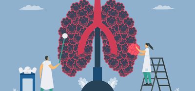
How 3D tissue shapes emerge in animal development
Posted: 14 August 2024 | Drug Target Review | No comments yet
Researchers showed that internal stress, induced by active cell behaviours, shapes the Drosophila wing disc pouch during eversion.
A mechanism by which tissues are programmed to transition from a flat state into a three-dimensional (3D) shape has been discovered by scientists at the Max Planck Institute of Molecular Cell Biology and Genetics (MPI-CBG), the Excellence Cluster Physics of Life (PoL) at the TU Dresden, and the Center for Systems Biology Dresden (CSBD).
The scientists used Drosophila, studying its wing disc eversion, with a method they developed to measure 3D shape changes and subsequently analyse cell behaviour throughout this process. Their research indicates that the shape programming method may be a common way to demonstrate how animal tissues form.
The research groups of Carl Modes, group leader at the MPI-CBG and the CSBD, and Natalie Dye, group leader at PoL and previously affiliated with MPI-CBG, aimed to discover how this shape change happens. Dye commented: “To explain this process, we drew inspiration from “shape-programmable” inanimate material sheets, such as thin hydrogels, that can transform into three-dimensional shapes through internal stresses when stimulated.” She added: “This concept has already helped us understand how plants grow. Animal tissues, however, are more dynamic, with cells that change shape, size, and position.”
The Drosophila wing disc eversion, when the dome shape transforms into a curved fold, showed shape programming is a useful mechanism to understand animal development. Jana Fuhrmann, postdoctoral fellow in the research group of Dye, explained: “Using a physical model, we showed that collective, programmed cell behaviours are sufficient to create the shape changes seen in the wing disc pouch. This means that external forces from surrounding tissues are not needed, and cell rearrangements are the main driver of pouch shape change.”
By reducing cell movement, the researchers assessed whether rearranged cells are the main reason for pouch eversion. Reducing cell movement caused issues with the tissue shaping process. Abhijeet Krishna, a doctoral student in the group of Carl Modes at the time of the study, said: “Our models combine both types of effects and link them directly to cell behaviours.”
Dye and Modes concluded: “Overall, our work suggests that early mechanical signals help organise how cells behave, which later leads to changes in tissue shape. Our work illustrates principles that could be used more widely to better understand other tissue-shaping processes.”
This study was published in Science Advances.
Related topics
Cell Therapy, Regenerative Medicine
Related organisations
Center for Systems Biology Dresden (CSBD), Excellence Cluster Physics of Life (PoL), Max Planck Institute of Molecular Cell Biology and Genetics (MPI-CBG), TU Dresden
Related people
Carl Modes (MPI-CBG and CSBD) ), Natalie Dye (PoL)