MRI technique differentiates benign breast lesions from malignancies
Posted: 20 February 2018 | Dr Zara Kassam (Drug Target Review) | No comments yet
An MRI technique that requires no contrast agent, combined with sophisticated data analysis, could reduce the number of unnecessary breast biopsies…
An MRI breast imaging technique that requires no contrast agent, combined with sophisticated data analysis, could reduce the number of unnecessary breast biopsies, according to a new study.
Researchers recently studied an alternative approach that eliminates the need for contrast agents in some cases by using diffusion-weighted imaging (DWI) measurements derived from MRI. The technique, known as diffusion kurtosis imaging, provides a picture of breast tissue on a microstructural level.
“Diffusion kurtosis imaging has been introduced in DWI to provide important information on tissue structures at a microscopic level,” said study lead author Dr Sebastian Bickelhaupt, from the German Cancer Research Center in Heidelberg, Germany. “Since malignant lesions disrupt the tissue structures at this level, diffusion kurtosis might serve as a relevant marker of changes.”
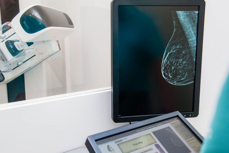
Dr Bickelhaupt, co-lead author Paul Jaeger, and colleagues evaluated a retrospective analysis of data collected from 222 women at two independent study sites. The women had suspicious findings on mammography that were classified under the Breast Imaging Reporting and Data System (BI-RADS) as BI-RADS 4 and 5 breast lesions. A BI-RADS 4 lesion is considered a suspicious abnormality, while a 5 is considered highly suspicious of malignancy. The women underwent DWI followed by a biopsy.
For the analysis, a software algorithm was developed for lesion characterisation, and imaging features were extracted using a kurtosis-based radiomics model. Radiomics is a rapidly growing field that enables the extraction of a large amount of quantifiable data from images.
In an independent test set of 127 women, the radiomics analysis reduced false-positive findings by 70 percent, while detecting 60 of 61 malignant lesions, or 98 percent.
“The model might help to lower the number of BI-RADS 4 lesions suspected of being cancer on the basis of screening mammography while retaining a high sensitivity similar to the sensitivity reported for biopsies themselves,” Mr Jaeger said.
Should the results hold in larger trials, the model has potential advantages in the clinic beyond its ability to reduce unnecessary biopsies in women with BI-RADS 4 lesions. The software algorithm makes the assessment reader-independent, ensuring that its accuracy is maintained across different imaging facilities.
The new approach is not intended to replace current contrast-enhanced breast MRI protocols in general, Dr Bickelhaupt emphasised, but to expand the spectrum of options available for answering specific clinical questions.
“This might also improve the efficiency of reporting,” he said.
The study appears online in the journal Radiology.
Related topics
Imaging, Label-Free, Magnetic resonance images (MRI), Oncology
Related conditions
Breast cancer
Related organisations
German Cancer Research Center
Related people
Dr Sebastian Bickelhaupt, Paul Jaeger