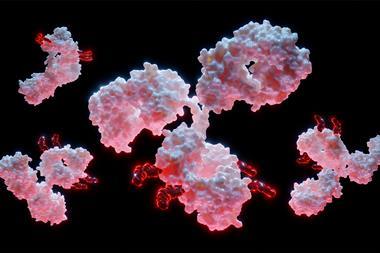
Despite significant strides in cancer research, metastasis remains the primary cause of cancer-related mortality globally. Joby Chesnick and Tracey Long, from the Digital Biology Group and Life Science Group, respectively, at BioRad Laboratories, reveal how investigation into circulating tumour cells (CTCs) and other rare cell types has led to the development of liquid biopsies as a potential new avenue for cancer diagnosis and monitoring.
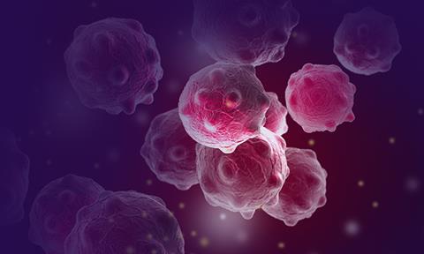
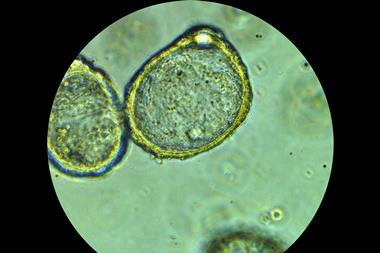
Cancer metastasis is thought to involve endothelial-mesenchymal transition (EMT) in a sub-population of solid tumour cells. During this process, a change in tumour cell gene expression profiles occurs which results in these cells becoming non-adherent and capable of entering the surrounding interstitial fluid. From there, CTCs enter the bloodstream and lymphatic system and travel to distant locations in the body. Migrated CTCs therefore have been proposed to contribute to the establishment of secondary tumours. The characterisation of CTCs from blood samples (ie, liquid biopsy) aimed at discerning the mechanisms of metastasis has gained significant attention as a cancer research focus.
Key considerations for antibody selection and preparation
The development of sophisticated single-cell technologies has driven recent interest in the enumeration and analysis of CTCs, shed by the primary tumour through the bloodstream and lymphatic system.1 Enumeration of captured CTCs and other rare cell types demands highly sensitive and robust techniques. As such, a critical aspect of successful cell enumeration lies in the design of the antibody panel. This process involves selecting appropriate antibodies and subsequently determining the optimal antibody concentration.2 An optimised staining panel must be created following careful planning and consideration of key panel components:
- Target cell:
Clearly defining the target cell type is crucial for the success of single-cell analysis. Researchers must decide between epithelial and mesenchymal cells based on the cancer type of interest. For instance, epithelial cells are prevalent in breast, colon and prostate cancers, while mesenchymal cells are characteristic of sarcomas. For example, MCF-7 epithelial breast cancer cells are derived from adenocarcinoma of the mammary glands.
- Target antigens:
Identifying the appropriate antigens expressed on the target cell is essential for positive identification. Researchers must consider both surface and intracellular antigens, as well as those not expressed by the target cell. This comprehensive approach allows for confirmation of identity and enhances the specificity of the staining panel.
- Fluorophore selection:
Careful selection of fluorophores is critical for the successful visualisation of target cells. Bright and highly photostable fluorophores with low susceptibility to photobleaching should be selected. Avoiding fluorophores prone to photobleaching, such as fluorescein isothiocyanate (FITC) and R-phycoerythrin (RPE), is advised. Additionally, researchers must choose fluorophores with non-overlapping emission spectra, especially when using multiple secondary antibodies. Finally, configuration of the fluorescent microscope should be considered, to ensure that the available filters are suitable for the selected fluorophores.
Once the fluorophore has been selected, researchers must then determine which to use for each antigen. The brightest fluorophore should typically be used to detect the antigen with lowest expression levels, and vice versa.
- Antibody staining method and antibody selection:
Using direct, indirect or a combination of both staining methods impacts the visualisation of target cells. In direct staining, a fluorescently conjugated primary antibody binds directly to the target antigen. Conversely, indirect staining employs an unconjugated primary antibody, followed by a fluorescently conjugated secondary antibody, enabling signal amplification.
Incorporating both staining methods simultaneously offers a comprehensive approach that can optimise detection sensitivity and specificity for a wide range of antigens. This dual approach facilitates the enumeration of CTCs with varying antigen expression levels, enhancing the overall detection capability of the panel.
The selection of primary antibodies is a crucial step in identifying the target cell. Monoclonal antibodies are recommended due to their high specificity for individual regions of the target antigen, unlike polyclonal antibodies, which may lead to increased background signal. The choice of primary antibody depends on the staining method used, the target species, antigen and conjugated format. In the case of MCF-7 cells, an antibody with specificity for human cells and cytokeratin (CK) is suitable.
In indirect staining, choosing a secondary antibody that targets the isotype of the primary antibody is essential. For instance, if the primary antibody is of IgG2a isotype, the secondary antibody must specifically detect IgG2a. Additionally, the conjugated format of the secondary antibody should be carefully chosen. An example is Goat Anti-Mouse IgG2a, suitable for MCF-7 cells stained with Mouse Anti-Human CD45.
Determining the optimal concentration of both primary and, if applicable, secondary antibodies is crucial to avoid nonspecific background staining or loss of signal intensity. Researchers must perform titration for each antibody to identify the appropriate concentration for target cell identification. This step is essential for the success of downstream single-cell analysis, ensuring accurate and reliable results.
Antibody titration and experiment setup
As a starting point for antibody titration, researchers can refer to the recommended dilution for immunofluorescence (IF) specified on the product datasheet provided by the antibody supplier. In cases where a recommended dilution is not available, the initial dilution for titration can be determined through an IF experiment. Throughout the process of identifying the optimal antibody concentration and configuring the experiment, some key steps need to be followed to ensure successful assay setup:
- Sequential titration: conduct only one concentration of the titration at any given time, commencing with the supplier’s recommended starting dilution.
- Inclusion of controls; incorporate positive and negative controls in each experiment. It is essential to titrate antibodies for negative controls to ensure accuracy. For example, false-positives can be ruled out by staining with the secondary antibody alone.
- Utilising representative cell lines: when determining the optimum antibody concentration, employ a representative cell line known to express the target antigen.
- Adjusting dilution: if the signal intensity is either too high or too low, repeat the experiment using the same antibody at a different dilution.
- Enumeration protocol: execute the direct or indirect enumeration protocol for each tested antibody. Strict adherence to the provided guidelines is imperative for valid results. Any deviation may compromise the integrity of the obtained data.
The analysis of CTCs and other rare cells must overcome various challenges to achieve widespread clinical adoption, including low abundance in blood, contamination with leukocytes, loss of cell viability during purification, and the involvement of labour-intensive, low-throughput processes.5 Careful panel design, use of online tools, and innovations in single-cell technologies offer automated easy-to-use, robust and precise approaches to CTC enumeration and analysis that has the potential to implement and accelerate the implementation of CTCs as a biomarker in cancer diagnosis.
About the authors
Joby Chesnick
Senior Segment Marketing Manager in the Digital Biology Group at Bio-Rad Laboratories
Joby manages marketing and communications for the Single Cell product portfolio. She earned BS and MS degrees in biology at Western Illinois University, followed by a PhD in plant biology at Texas A&M University. Dr Chesnick went on to receive a National Science Foundation Post-Doctoral Fellowship in Plant Biology while at the University of Washington where she characterised the chloroplast genome of an algal (dinoflagellate & diatom) endosymbiosis to elucidate the mechanisms of early organelle biogenesis. She has since held a variety of positions in both the biotech and molecular diagnostics industries, earned an MBA at the University of Wisconsin, and most recently joined Bio-Rad Laboratories in 2022 in her present role.
Tracey Long
Senior Marketing Manager in the Flow and Antibodies Group at Bio-Rad Laboratories, Inc.
Tracey and her team manage the product development and marketing for the catalog antibodies and StarBright™ Dyes portfolio. Tracey graduated from the University of Birmingham with a BSc (Hons) in biological sciences followed by a PhD in cellular and molecular virology, being awarded an MRC scholarship. Her thesis research focused on the development of vaccine candidates for Cytomegalovirus. Tracey has worked in business development, product management and marketing in both genomic and proteomic biotech, life science and healthcare companies, joining Bio-Rad in 2014 as a Product Manager and subsequently being promoted to Senior Marketing Manager.
References:
- Circulating Tumor Cell (CTC) Enumeration Overcomes Limitations of other Blood-Based Cancer Detection Methods. Bulletin 3267. Available from: https://www.bio-rad.com/sites/default/files/2021-12/DOC007-R10-Enumeration-and-Enrichment-App-Note-24NOV2021.pdf
- Antibody Selection and Preparation for Circulating Tumor Cell (CTC) and Rare Cell Isolation Using the Celselect Slides Enumeration Assay. Bulletin 3602. Available from: https://www.bio-rad.com/sites/default/files/2024-01/Bulletin_3602-Celselect-validated-antibody-quick-guide.pdf
- Interactive Human Immune Cell Marker Database. Available from: https://www.bio-rad-antibodies.com/human-immune-cell-marker-expression-antibodies-poster-guide.html
- Fluorescence SpectraviewerAvailable from: https://www.bio-rad-antibodies.com/spectraviewer.html
- Ring A, Nguyen-Sträuli BD, Wicki A, Aceto N. Biology, vulnerabilities and clinical applications of circulating tumour cells. Nat Rev Cancer. 2023;23(2):95-111. Available from: https://www.ncbi.nlm.nih.gov/pmc/articles/PMC9734934/