



Oligonucleotides can remain in tissues long after disappearing from circulation, but does accumulation necessarily indicate toxicity? Dr Tina Rogers examines the evidence needed to interpret preclinical safety findings.
Oligonucleotide therapeutics (oligos) present a distinct set of challenges for preclinical safety assessment. Unlike many small molecules, where plasma exposure can provide a useful approximation of systemic drug levels, oligos may disappear relatively quickly from circulation yet remain far longer in tissues. As a result, understanding where an oligo accumulates, how long it remains and its excretion route is essential for interpreting toxicology findings.
Biodistribution is therefore more than a descriptive pharmacokinetic (PK) exercise. The level of tissue exposure can be an important part of the safety assessment, particularly when findings occur in organs such as the liver or kidneys, where many oligos can reach high concentrations.
However, accumulation alone does not necessarily determine toxicity. Safety data may originate from several overlapping mechanisms, including chemistry-driven protein interactions, tissue exposure, hybridisation-dependent off-target activity and sequence-specific effects. The central challenge is determining what drives the biological response when an oligo reaches a particular tissue.
What are oligonucleotide therapeutics?
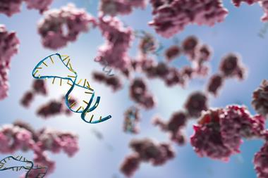
Oligonucleotide therapeutics, often referred to as oligos, are medicines made from short, synthetic strands of DNA or RNA, or chemically modified versions of these molecules. They are designed to act on specific genetic sequences, often by binding to RNA to alter gene expression or protein production.
Examples include antisense oligonucleotides (ASOs) and small interfering RNAs (siRNAs). Unlike many conventional small-molecule drugs, oligos can remain in tissues long after their concentrations in the blood have declined. Understanding where they accumulate and how long they persist is therefore important when assessing their safety.
Why plasma exposure is only part of the story
One of the defining characteristics many oligos share is the disconnect between plasma and tissue kinetics. Plasma half-life can be relatively short, yet the drug remains in tissue for days or weeks, thus plasma concentrations may not fully represent the magnitude or duration of intended or off-target exposure.
The distinction becomes particularly important during repeat-dose studies. Accumulation may continue as additional doses are administered before material from previous doses fully clears. This also means plasma concentration may provide incomplete histopathologic or clinical pathology data.
Interpreting oligonucleotide safety can benefit from measurements in both plasma and relevant tissues when feasible. Tissue Cmax, the highest measured concentration of a substance in a tissue, and area under the curve (AUC), which reflects overall tissue exposure over time, can help place toxicology findings in the context of both the magnitude and duration of local exposure. These measurements may be informative in target organs, tissues associated with uptake or clearance and organs in which treatment-related findings are observed.
Conjugated oligos are chemically linked to a targeting or delivery moiety, whereas unconjugated oligos lack this additional component. In conjugated programmes, tissue exposure data may need to be interpreted alongside information on conjugate stability and cleavage. Measuring the intact conjugate and the released or free oligonucleotide can help clarify which molecular species is contributing to the observed tissue Cmax and AUC, and which form may be most relevant to a safety finding.
Integrating these approaches should not replace conventional toxicokinetic measurements. They are intended to help interpret systemic exposure with the drug concentrations present at the site of an observed effect.
Accumulation does not automatically equal toxicity
High concentration of an oligo in a tissue is not, by itself, evidence of toxicity. Oligos can accumulate in highly perfused organs and tissues involved in uptake and clearance, such as the liver and kidneys. The spleen and lymph nodes are also common areas of drug concentration, depending on the molecule and its absorption, distribution, metabolism and excretion (ADME) characteristics.
Whether that accumulation becomes biologically meaningful depends on local concentration, chemistry and sequence. High tissue exposure may coincide with a problematic safety finding without necessarily being its sole cause – ie, a correlation between exposure and pathology does not establish mechanism. An unexpected observation in the liver or kidney, for example, may be associated with local accumulation, but chemistry-dependent interactions or sequence-related activity may also contribute.
Tissue exposure is therefore most informative when interpreted as part of a broader evidence package rather than as a standalone endpoint.
Multiple endpoints can converge to generate an unexpected safety finding
A key oligo toxicology challenge happens when several potential drivers of toxicity operate simultaneously:
- Chemistry-driven effects may arise from the physicochemical characteristics of an oligonucleotide and its interactions with proteins. These effects may be influenced by features such as backbone chemistry, modifications, length or conjugation.
- High and sustained tissue accumulation may contribute to organ-specific findings, particularly in the liver and kidney.
- Oligos may interact with unintended RNA sequences, leading to off-target effects. These effects are inherently more sequence-dependent and require a different approach to investigation than chemistry-driven findings.
- Individual sequences may modify the safety profile even within a common chemical platform.
These endpoints are not mutually exclusive. A finding observed in a highly exposed organ could reflect a combination of tissue concentration, chemical characteristics and sequence-dependent biological activity. The development challenge is separating correlation from causation as far as the available data allow.
Building an integrated evidence package
No single assay is likely to explain every unexpected toxicology finding. A more robust approach is to integrate information across several complementary data streams.
1. Tissue exposure and biodistribution can establish where the oligonucleotide accumulates, the magnitude of that exposure and how long it persists
2. Toxicokinetic data can help place tissue findings in the context of dose and systemic exposure, including changes that emerge after repeated administration
3. Clinical pathology and biomarkers can provide evidence of functional changes associated with organ or class-related effects
4. Histopathology can define the location and character of tissue changes and allow those observations to be compared with measured tissue concentrations
5. Off-target assessment can help investigate whether sequence-dependent hybridisation may be contributing to a finding that cannot be explained adequately by exposure or chemistry alone.
Together, these data can help development teams ask more informative questions. Does the finding occur in the tissue with the greatest exposure? Does severity increase as tissue concentrations increase? Does the finding persist after plasma exposure has fallen? Is the pattern consistent with a broader chemistry-related effect, or is there evidence suggesting sequence-specific activity? Interpreting these datasets together is where the true value lies.
Connecting biodistribution with histopathology
One particularly useful application of this integrated approach is in studying the relationship between tissue concentrations, histopathology and clinical pathology. Quantitative biodistribution studies can include major organs, blood or plasma and the administration site, with sampling designed to capture absorption, peak tissue levels and elimination. These measurements can be compared with the timing and severity of observed tissue changes.
For oligos, particular attention may be warranted for the liver, kidney, spleen, bone marrow and lymph nodes, depending on the molecule and study design. This type of analysis can help determine whether pathology reflects local exposure and whether an apparent recovery in clinical pathology corresponds with declining tissue concentrations. It can also reveal situations in which tissue levels remain substantial even after circulating concentrations have fallen.
Applied this way, biodistribution becomes directly relevant to toxicological interpretation rather than serving solely as a characterisation of drug disposition.
Tissue persistence can also influence study design
Understanding accumulation is important for both interpreting completed studies and for designing subsequent ones. Long tissue residence can influence the duration of recovery periods. If an oligo remains present in an organ long after the end of dosing, a recovery interval that is too short may provide limited information about whether a finding is reversible. Persistent tissue exposure associated with ongoing toxicity – and insufficient recovery data to characterise that toxicity – can both complicate dose selection and clinical monitoring, potentially increasing risk to patients.
The same principle can inform planning for repeat-dose and longer-duration studies. Evidence of accumulation can help determine whether additional monitoring is warranted and whether the timing of tissue collection adequately captures both peak exposure and elimination.
Biodistribution helps answer related questions about what the findings mean immediately and how subsequent studies should be designed to better understand the implications.
From distribution data to safety interpretation
Optimal evaluation of oligo programmes goes beyond answering, “Where does the molecule go?” They should explore how exposure to specific tissues influences biological observations. This requires a risk-based, chemistry- and target-informed approach that integrates bioanalysis, toxicokinetics, biodistribution, pathology, biomarkers and off-target assessment.
Tissue accumulation is an important part of that picture, but it should not be interpreted in isolation. By examining exposure alongside the other potential drivers of toxicity, development teams can build a more complete explanation for observed findings and use that understanding to inform recovery assessments, subsequent study design and the broader preclinical safety strategy.








No comments yet