


New research from MIT demonstrates that cell membranes play an active role in regulating epidermal growth factor receptor (EGFR) activity. Using nanodiscs and single molecule FRET, researchers found that high levels of negatively charged lipids lock EGFR into a constitutively active state, whilst cholesterol suppresses signalling, offering potential new therapeutic strategies targeting membrane composition in aggressive cancers.
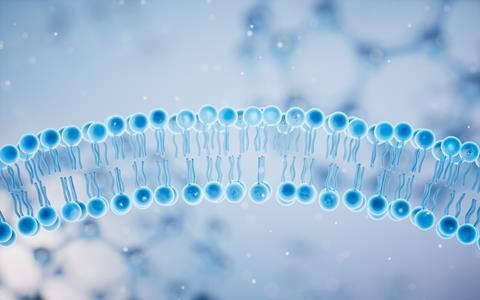
Cell membranes have long been viewed as little more than protective barriers that provide structure and separate cells from their surroundings. However, research from the Massachusetts Institute of Technology (MIT) is adding to growing evidence that these membranes may play a much more active role in controlling cellular behaviour.
In the study, MIT researchers found that changes in the composition of the cell membrane can significantly affect the activity of epidermal growth factor receptor (EGFR), a protein known to drive cell growth and proliferation.
The findings could help explain why certain cancer cells become highly aggressive and may eventually lead to new therapeutic approaches aimed at regulating receptor activity through the membrane itself.
“The longstanding dogma of what a membrane does is that it’s just a scaffold, an organisational structure. However, there have been increasing observations that suggest that maybe these membrane lipids are actually playing a role in receptor function,” says Gabriela Schlau-Cohen, the Robert T. Haslam and Bradley Dewey Professor of Chemistry at MIT and senior author of the study.
Investigating receptor behaviour
EGFR is found on the surface of cells that line organs and body surfaces and is key in regulating growth. Overexpression of the receptor has been linked to several cancers, including lung cancer and glioblastoma, where excessive signalling can lead to uncontrolled cell division.
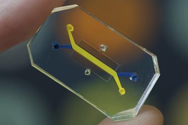
Because EGFR spans the entire cell membrane, understanding how signals are transmitted through the protein has been a challenge for researchers. To overcome this, the MIT team used nanodiscs, self-assembling membrane structures that closely mimic natural cell membranes while allowing researchers to study the full-length receptor in a controlled environment.
EGFR is found on the surface of cells that line organs and body surfaces and is key in regulating growth
The researchers combined these nanodiscs with a technique known as single molecule fluorescence resonance energy transfer (FRET), which enabled them to observe how the receptor changes shape under different membrane conditions.
Previous work by the team had shown that when EGFR binds to epidermal growth factor (EGF), the receptor undergoes a structural change that activates growth-promoting pathways inside the cell.
Researchers interested in membrane protein analysis can learn more in the webinar From expert task to lab routine – membrane protein purification and analysis simplified, which explores automated workflows and technologies designed to simplify membrane protein purification and stability analysis while preserving native protein structure.
High levels of negatively charged lipids lock EGFR into growth mode
In the latest study, the researchers investigated how altering the membrane’s lipid composition affected EGFR activity.
Under normal conditions, around 15 percent of the cell membrane consists of negatively charged lipids. The team found that membranes containing between 15 and 30 percent of these lipids functioned normally. However, when levels reached 60 percent, EGFR became locked into an active state.
Under normal conditions, around 15 percent of the cell membrane consists of negatively charged lipids
In this configuration, growth-promoting signals remained switched on continuously, even in the absence of EGF.
Many cancer cells are known to contain elevated levels of negatively charged lipids, suggesting this mechanism could contribute to their unchecked growth.
“If the membrane has high levels of negatively charged lipids, then it’s always in that open conformation. It doesn’t matter if ligand is bound or unbound,” says Schlau-Cohen. “It’s always in the conformation that’s telling the cell to grow, not just when EGF binds.”
Cholesterol shown to have opposite effect
The team also examined the influence of cholesterol on receptor behaviour. When cholesterol levels were increased within the nanodiscs, the membranes became more rigid and EGFR signalling was suppressed.
The results provide further evidence that membrane composition can directly regulate receptor activity and may offer new opportunities for cancer treatment.
According to the researchers, future therapies could potentially target membrane properties themselves, including strategies designed to neutralise excessive negative charge and reduce EGFR signalling in tumour cells.
The study adds to a growing body of research suggesting that cell membranes are not merely passive structures but active participants in the molecular processes that control health and disease.


No comments yet