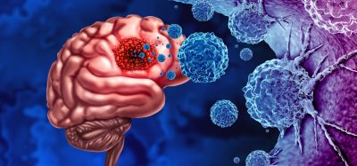
Muscle cryomicroscopy images could provide model for nanomachines
Posted: 9 March 2017 | Osaka University | No comments yet
Myosin provides model for nanomachines
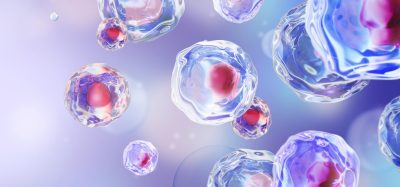
High-resolution cryomicroscopy images have provided new insights into how myosin generates force in muscle contraction and a model for the construction of nanomachines.
The results, published in Nature Communications, revealed unexpectedly large conformational changes in the myosin molecule during muscular contraction. Professor Keiichi Namba, of Osaka University, where the research was conducted, said: “Myosin and actin are nanomachines that convert the chemical energy of ATP hydrolysis into mechanical work.”
Myosin converts this energy by hydrolysing ATP molecules into movement along an actin filament. The hydrolysis involves several conformational changes in myosin. These changes have been imaged using electron microscopy, but there were previously no atomic images of ATP hydrolysis when myosin is interacting with actin, which would more accurately represent the changes in myosin during muscle contraction.
Electron cryomicroscopy images at 5.2 Å resolution showed a previously unobserved conformational change in the myosin molecule when it interacts with actin. The researchers hypothesised that this conformation could explain why muscle myosin has far faster kinetics than other myosin in the body.
Prof Namba believes that the structural information gained by this work could be used to make artificial nanomachines. “We are studying nature’s nanomachines to build man-made ones,” he said.
Related topics
Microscopy
Related organisations
Osaka University
Related people
Professor Keiichi Namba