During embryogenesis, organs must achieve a precise cell count and functional structure. Embryos efficiently manage multiple tasks—growth, shaping, and architecture establishment. Despite extensive research on embryonic development, the intricate coordination of these tasks in both space and time remains incompletely understood.
Addressing this question, Caren Norden (group leader) and Mauricio Rocha-Martins (postdoctoral researcher) conducted a study involving a multidisciplinary team, including computer scientists. They employed advanced technology to explore how the vertebrate retina adapts to the challenges of simultaneous growth and tissue remodelling.
Unique model systems
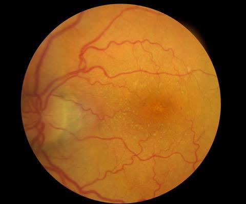
The study focused on zebrafish embryo retinas and human retinal organoids. These systems possess distinct advantages due to their small size and translucency, enabling real-time observation of tissue organisation and growth. State-of-the-art techniques such as light-sheet microscopy and deep learning-based image restoration provided unprecedented insights into cellular behaviours.
Neuronal relocation strategy
Researchers made a fascinating observation: a group of neurons called photoreceptors temporarily shifts away from their typical tissue zone. This relocation creates space for progenitor cells to divide, generating additional cells that contribute to the developing neuronal retina. When photoreceptor movement is hindered, congestion occurs, compelling progenitor cells to divide in incorrect regions, leading to tissue malformation. Therefore, this temporary migration of neurons prevents interference with progenitor cells, promoting the harmonious development of the organ.
Novel insights and implications
Mauricio Rocha-Martins, the study's lead author, noted that this migration phenomenon showcases that neuronal migration does not solely involve reaching the correct location. It can also directly influence the coordination of organ development. The significance of these findings extends beyond retinal development. The simultaneous growth and establishment of functional architecture are common features in various developing organs. The results open avenues to explore similar strategies in other developing organs. Furthermore, considering that defective neuronal migration contributes to severe brain malformations in humans, these findings emphasize the need to understand cell interactions comprehensively to unravel the causes of developmental disorders.