Artificial intelligence cell analysis for swift blood disease diagnosis
Posted: 17 August 2023 | Drug Target Review | No comments yet
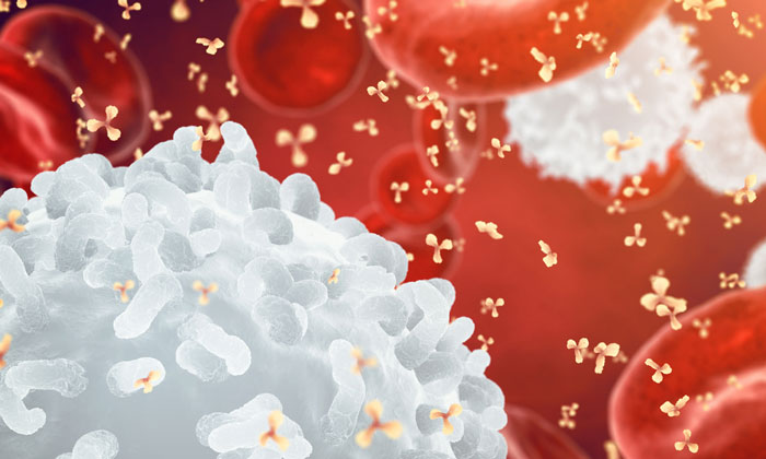
Researchers from the German Cancer Research Center (DKFZ) and the Cambridge Stem Cell Institute have developed an artificial intelligence (AI) system capable of identifying and characterising white and red blood cells within microscopic images of blood samples. This AI algorithm holds the potential to aid medical professionals in diagnosing blood disorders and is openly accessible for research purposes.


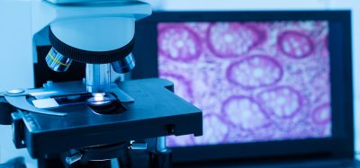
Blood-related ailments are frequently characterised by irregular quantities and unusual shapes of both red and white blood cells. Traditional diagnosis of these conditions involves the manual examination of blood smears under a microscope. While this method is straightforward, it can prove challenging for experienced experts to evaluate subtle alterations that may affect only a small subset of the numerous visible cells.
As a result of these challenges, distinguishing between different diseases is not always straightforward. For instance, observable changes in the blood of patients with myelodysplastic syndrome (MDS), an early form of leukaemia, often resemble those found in less severe cases of anaemia. Consequently, a definitive diagnosis of MDS necessitates more invasive procedures like bone marrow biopsy analysis and molecular genetic testing.
“To assist specialists in navigating these intricate diagnoses, we’ve devised a computer-based system that automatically identifies and characterises white and red blood cells present in peripheral blood,” explains Moritz Gerstung of DKFZ. Gerstung and his team initially trained the algorithm, known as Haemorasis, using over five hundred thousand instances of white blood cells and millions of red blood cells from more than three hundred individuals with diverse blood disorders (including various types of anaemias and forms of MDS).
Gerstung notes, “The algorithm is proficient at detecting the shapes and quantities of tens of thousands of blood cells within a microscopic blood image. This complements human capabilities, which usually tend to focus on finer details.” With this acquired knowledge, Haemorasis can now propose diagnoses for blood disorders and even differentiate between genetic subtypes of these conditions. Furthermore, the algorithm unveils specific associations between particular cell structures and diseases, a task that is often intricate due to the large number of cells involved.
Published in Nature Communications, Haemorasis has already undergone testing on three separate patient groups to validate its functionality across various testing facilities and blood count scanning systems. “For the first time, we have shown that computer-assisted analysis of blood images is viable and can contribute to initial diagnostics,” Gerstung states. Haemorasis is tailored to enhance hematology diagnostics, thereby aiding in more precise initial diagnoses of blood-related disorders. This is particularly valuable in identifying patients necessitating further invasive procedures such as bone marrow biopsies or genetic analyses.
“In the future, automated cell analysis using Haemorasis could complement routine blood disorder diagnoses. Although the algorithm has currently been trained on specific diseases, we envision substantial potential in this approach,” Gerstung emphasises. He underscores the need for additional studies to uncover any potential limitations of the method.
Related topics
Artificial Intelligence, Bioinformatics
Related conditions
blood disorders
Related organisations
Cambridge Stem Cell Institute, German Cancer Research Center (DKFZ)
Related people
Moritz Gerstung (German Cancer Research Center)


