New technique to extract and analyse extracellular vesicles
Posted: 8 November 2023 | Drug Target Review | No comments yet
Cellulose nanofiber sheets enable analysis of EVs, and miRNAs within them, offering potential for cancer treatment and personalised medicine.
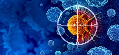
A technique using cellulose nanofiber (CNF) sheets derived from wood cellulose has been developed by Nagoya University scientists to capture extracellular vesicles (EVs) from fluid samples and organs during surgery. EVs are small structures from cancerous cells that are essential for cell-to-cell communication. Extracting and analysing EVs using this new technology could potentially revolutionise early cancer diagnosis and personalised medicine.
Many cancers have poor prognosis and go undetected until its advanced stages, leaving patients with limited treatment options. Using EVs enables physicians to find the cancer early and get essential information on disease status and its progression, which enables them to create personalised cancer treatment plans. However, researchers have found it difficult to use EVs because of the lack of an effective isolation strategy.
Capturing EVs
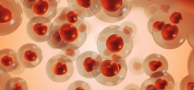
The team, led by Dr Akira Yokoi, used CNF sheets made from wood cellulose to extract EVs from fluid samples from ovarian cancer mice models. The sheet, which has a porous nanostructure, absorbs the fluid containing EVs into its pores and closes them when it dries. The sheets captured and preserved EVs from ten microliters of body fluids. The current standard methods, like ultra-centrifugation, are more time-consuming and require much larger samples in contrast.
Dr Yokoi said: “We have developed the unique cellulose nanofiber by applying paper-making and solvent displacement technology.” Yokoi continued: “The cellulose nanofibers we use are a sustainable biomass material that comes mostly from wood cell walls. These sheets have attractive properties, such as being lightweight, high strength, and most importantly, easily biodegradable.”
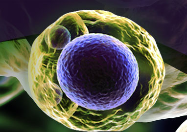
This technique enabled the scientists to successfully extract and analyse EVs, and the microRNAs (miRNAs) contained within them from the mice ovarian cancer models. Healthy patients’ miRNAs differ from sick patients’ miRNAs, so they make an ideal diagnostic marker for cancer.
The team also found distinct sets of miRNAs in EVs collected from tumour surfaces, some of which decreased after tumour removal. Monitoring the presence or absence of these miRNAs may be a straightforward way to analyse the effectiveness of treatment and then adapt treatment according to tumour heterogeneity. Heterogeneity is a frequent issue where even in a single tumour, cancer cells have distinctive characteristics and properties.
The sheets have a similar structure to medical gauze, so they can be attached and removed easily, even when put on organs during surgery. The researchers used recently removed human organs to test this and were successful, finding that the EVs on the tumour surface showed unique miRNA profiles compared to the tumour tissue.
Personalised medicine
“Organ surfaces were a previously unanalysed EV subpopulation, which can now be subjected to biological assessments,” Yokoi explained. “CNF paper enables the obtaining of EVs from multiple sites in the body. Then, by checking the molecular profiles of these EVs, we can monitor disease progression and tailor the selection of the best drug, contributing to personalised medicine.”
Dr Takahiro Ochiya, Board Member of the International Society for Extracellular Vesicles and President of the Japanese Society for Extracellular Vesicles said: “Exosome analysis using CNF sheets is an extremely novel method and is expected to have a variety of applications, including medical uses. We expect this to be a major advance that will bring the knowledge of exosomes as medical research directly to patients.”
This research has many implications, pioneering the analysis of EVs during surgery which has previously been unexplored. The team hopes to advance the medical application of EV sheets for various diseases, improve diagnostic accuracy, and help progress personalised medicine.
The study is published in Nature Communications.
Related topics
Cancer research, Personalised Medicine
Related conditions
Ovarian cancer