Researchers have developed a system that enabled quantitative assessment of cytotoxicity of SiNPs in human hepatoma cells.
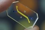
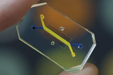
Scientists from the Graduate School of Pharmaceutical Sciences at Chiba University have estimated the cellular intake of silica nanoparticles (SiNPs) based on their sizes. They developed an AF4-ICP-MS (asymmetric flow field flow fractionation with inductively coupled plasma mass spectrometry) system, which separated SiNPs of five different sizes and enabled quantitative assessment of cytotoxicity of SiNPs in HepG2 cells.
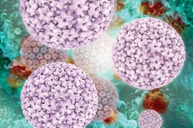
SiNPs
Silica nanoparticles exhibit excellent physicochemical properties, resulting in their wide acceptance in biomedical applications. However, there is a growing concern regarding its negative effects on human health. The nano-sized particles have shown high reactivity towards biomolecules and often toxicity towards biological cells.
This behaviour of metal nanoparticles is attributed to interaction with biomolecules to phenomena like inflammation or oxidative stress. To ensure the safe usage of metal nanoparticles, there is a need to explore molecular mechanisms responsible for the toxicity and understand how the uptake of nanoparticles by cells varies based on their shape, size, morphology, and other aspects. Therefore, the Chiba University team have proposed a new technique that can separate nanoparticles of different sizes, assess their uptake by cells, and evaluate the potential toxicity.
Therefore, the researchers adopted the novel AF4-ICP-MS size analysis technique, which overcame issues posed by size analysis techniques like electron microscopy and laser-based dynamic light scattering, which failed to observe nanoparticle specimens in deep layers and elucidate the chemical compositions of the nanoparticles. AF4-ICP-MS detected nanoparticles of 10, 30, 50, 70, and 100 nm.
The AF4-based method was used to evaluate the cellular uptake of SiNPs in lab-cultured human hepatoma HepG2 cells, with the measurements demonstrating that approximately 17 percent of the SiNPs exposed to the HepG2 cells were absorbed. Also, the team carried out transmission electron microscopy (TEM), which observed the presence of SiNP aggregates within the cells. This indicated the ability of small nanoparticles to settle in the culture medium and enter the cells easily.
Dr Yu-ki Tanaka, assistant professor, said of the toxicity experiments: “We found that the smaller SiNPs exhibited higher toxicity towards the HepG2 cells than the larger ones, but the AF4-ICP-MS analysis found no significant size-dependent difference in the particle volume absorbed by the cells.”
These findings indicated that the elevated cytotoxic behaviour of small SiNPs was because of the large surface area relative to particle volume.
Chemical mechanisms
Furthermore, the scientists explored the chemical mechanisms associated with cytotoxicity. The data showed that cell necrosis was partly linked to oxidative stress caused by the production of reactive oxygen species (ROS). Also, interactions of the silanol groups on the SiNP surface and phospholipids in the cell membrane were responsible for the associated cell damage.
The new AF4-ICP-MS technique is a powerful tool for quantitatively determining cytotoxicity induced by metal nanoparticles of varying sizes. The findings provide a basis for future studies evaluating nanoparticle exposure risks and their potential burden on human bodies.
Dr Tanaka stated: “We are hopeful that the toxicological information provided by our study will help establish criteria for the proper utilisation and regulation of nanoparticles in industries, the medical field, and even in daily use items containing nanoparticles.”
This study was published in Archives of Toxicology.