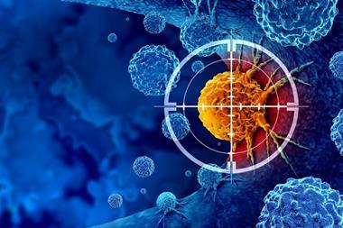
New imaging technology called fluorescence lifetime (FLT) imaging improves the accuracy of cancer surgery for multiple tumour types.
A team of scientists at Mass General Brigham in the US has developed a visualisation tool combining high-speed cameras, that can detect changes happening in a billionth of a second, and FDA-approved fluorescent injections to distinguish tumour tissue from normal tissue across several cancer types. The researchers reported that this technique was over 97 percent accurate across tumour types.
Distinguishing tumours
Dr Anand Kumar of the Athinoula A Martinos Center for Biomedical Imaging at Massachusetts General Hospital (MGH) said: “Our lab has been studying fluorescence lifetime imaging since 2002, but this is the first time that anyone has combined it with tumour imaging and injectable dyes in humans. By doing so, we’ve developed a technique for accurately distinguishing tumour tissue from healthy tissue across cancer types.”
“This technology has taken us to the brink of a revolution in solid tumour surgery.”
Kumar worked with colleagues from Mass Eye and Ear, another member of the MGH healthcare system, where head and neck cancer patients are treated. Dr Mark Varvares, chief of Otolaryngology-Head and Neck Surgery at Mass Eye and Ear, said: “This technology has taken us to the brink of a revolution in solid tumour surgery.” Revealing how it will improve treatment, he continued: “By using the advanced imaging techniques combined with the dye, surgeons in the near future will have the ability to more completely remove all malignant cells during tumour surgery while at the same time, with confidence, spare normal tissue, enhancing postoperative function and in some cases, the patient’s appearance.”
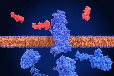
To improve the visualisation of tumours during surgery, many technologies have been pursued, such as fluorescence imaging and advanced microscopy. However, these technologies have not been broadly implemented and they are mostly restricted to specific types or subtypes of cancer. For example, fluorescence imaging uses dyes to target cancer-specific molecules, but standard imaging techniques provide limited accuracy for detecting tumour margins, the edges of normal tissue that surround a tumour, as the expression of these molecules greatly varies within and across tumour types.
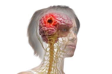
Complete surgical excision of tumours is a critical part of cancer treatment, to ensure optimum outcomes and lower rates of recurrence. Patients who suffer from osteosarcoma recurrence currently face less than 20 percent survival rate in the long term. While orthopaedic surgeons in oncology aim to salvage as much functional tissue as possible, they are often left with no other option than to resect beyond the tumour margin to decrease the risk of recurrence, which can often lead to amputation. Furthermore, after primary surgical treatment, patients face up to a 40 percent chance of cancer recurrence due to local relapse from residual marginal osteosarcoma cells that were not picked up initially by the surgeon.1
FLT imaging
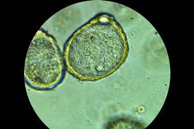
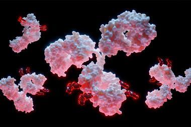
FLT imaging, adopted by Kumar and his associates, does not rely solely on dyes to target cancer. The technique also uses high-speed cameras to identify changes in the property of light emitted by tissue. In preclinical models from prior studies, the researchers found that tumours in mice injected with an indocyanine green (ICG) dye had a longer fluorescence lifetime compared to normal tissue. The difference allowed the researchers to precisely recognise normal tissue and tumour tissue.
In the current study, the team used the same principle for patient samples. They gave patients an ICG injection at least a day before their surgeries, and then analysed samples from liver surgery patients at MGH and head and neck patients at Mass Eye and Ear.
Following this, scientists from multiple institutions evaluated over 60 patient specimens, including brain, tongue, breast, liver, bone and soft tissue. These patients were treated at MGH, Mass Eye and Ear, University of Newcastle in the UK, Leiden University in the Netherlands, and The University of Pennsylvania.
The team detected an FLT shift at a cellular level that was consistent across tumour types and in different patients. It also differentiated between benign and metastatic lymph nodes.
Close to clinical impact
The researchers state that ICG is currently not approved by the FDA for clinical use as a tumour marking agent, so their next aim is to carry out a larger scale clinical trial to test FLT’s safety and efficacy with ICG for tumour identification during surgeries.
Kumar stated: “Our work suggests that the combination of fluorescence lifetime imaging with ICG could improve surgical resections, thereby impacting patient lives.” He added: “We’re excited to take these next steps to move our discoveries closer to clinical impact.”
The paper was published in Nature Biomedical Engineering.
Reference
1 Alakhras S, D’Acunto M, Ejaz M, et al. Surgical margin assessment of bone tumours: A systematic review of current and emerging technologies. Journal of Bone Oncology [Internet]. 2023 February 9 [2023 October 23]; 39:1-9. Available from: https://www.sciencedirect.com/science/article/pii/S2212137423000027