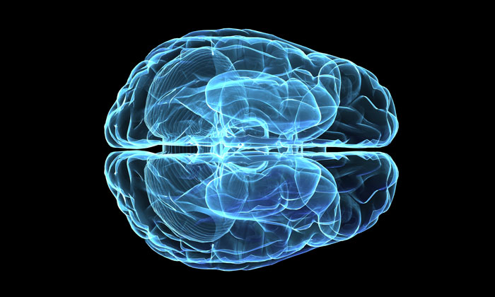
Raman imaging technique shows Alzheimer’s disease progress in the brain
Posted: 24 January 2018 | Drug Target Review | No comments yet
Using Raman optical technology, scientists can now produce images of brain tissue that is affected by Alzheimer’s disease…
Alzheimer’s disease is associated with areas of high protein concentration in brain tissue – plaques and tangles – and Raman imaging is now being used to obtain sharp images of these affected areas. It is an attractive technique, as it shows more than the specific proteins involved. The presence of water and lipids, influenced by protein presence, can also be detected. Using this technique, the researchers have studied brain tissue from four brain donors – three of them with Alzheimer’s disease.
Raman indicates transition
Raman application has enabled the affected area to be visible in a sharp and clear way. Furthermore, it also hightlights areas that are in transition between healthy and affected tissue, which may give an indication of how the disease is spreading in the brain. The studied brain tissue of the healthy person, also detected a small area with protein activity, which may indicate early signs of a neurodegenerative disease.
Raman microscopy uses a laser beam to detect chemical substances – the energy of the reflected and scattered light giving clear indication of the substances present in a sample – and in each of the four brain samples, 4096 spectra were examined in this way. A major advantage of Raman is that the chemicals don’t need a pretreatment; it is ‘label free’, which has proven Raman a useful technique in chemical analysis.
Cell level precision
In this case, Raman was used to examine brain tissue outside the body, but it could even be used ‘in vivo’ for detecting specific areas during surgery. Compared to MRI, PET and CT imaging, Raman is able to detect areas – even smaller than cells – with very high precision. In this way, it can be a highly valuable additional technique. The Raman images now show protein activity at neural cell level, but the sensitivity is high enough for detecting areas that are even smaller – as is the case with the brain sample of the healthy person.
Cees Otto, of the Medical Cell Biophysics group of UT, has published his work together with colleagues from Leiden University as well as from from Spain and Austria.
Related topics
Disease Research, Neurosciences, Personalised Medicine, Precision Medicine, Spectroscopy
Related conditions
Alzheimer’s disease
Related organisations
Leiden University, University of Twente
Related people
Cees Otto