Fragile X imaging study reveals differences in infant brains
Posted: 5 April 2018 | Dr Zara Kassam (Drug Target Review) | No comments yet
For the first time, researchers have used MRIs to show that babies with the neurodevelopmental condition fragile X syndrome had less-developed white matter compared to infants that did not develop the condition…
For the first time, researchers have used MRIs to show that babies with the neurodevelopmental condition fragile X syndrome had less-developed white matter compared to infants that did not develop the condition. Imaging various sections of white matter from different angles can help researchers focus on the underlying brain circuitry important for proper neuron communication.
The study shows that there are brain differences related to the neurodevelopmental disorder established well before a diagnosis is typically made at age three or later.
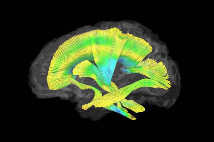
This image shows all of the white matter fibre tracts investigated in this study. Dr Meghan Swanson, UNC-Chapel Hill
“It’s our hope that earlier diagnosis and intervention will help children with fragile X and their families,” said co-first author Dr Meghan Swanson, a postdoctoral research fellow at the Carolina Institute for Developmental Disabilities at the UNC School of Medicine. “We also hope that this knowledge might inform drug development research.”
So far, drug clinical trials have failed to demonstrate a change in treatment targets in individuals with fragile X. One of the challenges has been identifying good treatment outcome measures or biomarkers that show the response to intervention.
“One of the exciting things about our findings is that the white matter differences we observe could be used as an objective marker for treatment effectiveness,” said co-senior author Dr Heather C. Hazlett, Assistant Professor of Psychiatry at the UNC School of Medicine.
For this study, Dr Swanson, Dr Hazlett, and colleagues imaged the brains of 27 infants who went on to be diagnosed with fragile X and 73 who did not develop the condition. The researchers focused on 19 white matter fibre tracts in the brain. Fibre tracts are bundles of myelinated axons – the long parts of neurons that extend across the brain or throughout the nervous system. Think of bundles of cables laid across the brain. These bundles of axons connect various parts of the brain so that neurons can rapidly communicate with each other. This communication is essential, especially for proper neurodevelopment during childhood.
Imaging and analytical analysis showed significant differences in the development of 12 of 19 fibre tracts in babies with fragile X from as early as six months of age. The babies who wound up being diagnosed with fragile X had significantly less-developed fibre tracts in various parts of the brain.
“These results substantiate what other researchers have shown in rodents – the essential role of fragile X gene expression on early development of white matter in babies,” said co-first author Dr Jason Wolff, a former postdoctoral fellow at UNC-Chapel Hill and now Assistant Professor of Educational Psychology at the University of Minnesota. “Our work highlights that white matter circuitry is a potentially promising and measurable target for early intervention. However, achieving the goal of infant intervention for fragile X would likely require expanded newborn screening efforts.”
The study has been published in JAMA Psychiatry.
Related topics
Biomarkers, Disease Research, Imaging, Magnetic resonance images (MRI)
Related conditions
Fragile x
Related organisations
UNC School of Medicine
Related people
Dr Heather C. Hazlett, Dr Jason Wolff, Dr Meghan Swanson