Featured content

Pomegranate-derived compound shows therapeutic potential in heart disease
Researchers at Cardiff University have identified urolithin A – a compound produced by gut bacteria during the metabolism of substances found in pomegranates – as a potential new approach for treating cardiovascular disease. Their findings suggest the molecule could help reduce inflammation and stabilise dangerous artery plaques, potentially lowering the risk of heart attacks and strokes.
 Article
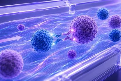
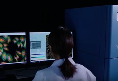
Flow-based human tumour models reveal immune responses missed by static culture
Static cultures may not tell the whole story when it comes to immunotherapy performance. Results from the Mera™ flow-based human tissue model show stronger T-cell activity and cytokine responses under physiological flow, highlighting the role of dynamic immune–tumour interactions in preclinical testing.
 Article
AACR 2026 part two: integrating AI, spatial biology and next-gen therapeutics
In part two of our AACR 2026 coverage, industry leaders were focussed on how the field is no longer constrained by data generation or molecular design, but by the challenge of connecting systems, standardising workflows and ensuring biological insights.
 Report
AI in Drug Discovery: Progress, Limits and What Comes Next
AI has attracted enormous investment across drug discovery, but major questions still remain around validation, reproducibility and real-world application. In our latest Beyond the Lab report, experts discuss where the technology is starting to influence discovery workflows – and where limitations continue to slow adoption.
Opinion and interviews
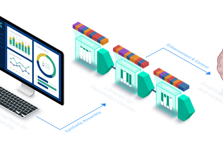
Computational design drives new generation of synthetic promoters
Designing gene control from scratch is becoming possible. SynGenSys is using computational design to create synthetic promoters for advanced therapies.
- Previous
- Next


Join now
Unlock exclusive industry insights
- Bookmark articles and resources to access anytime
- Enjoy free access to industry leading resources, webinars and insights
- Stay informed with the latest news and breakthroughs
- Receive updates and recommendations tailored to your research interests
Upcoming Webinars

Webinar
Where NAMs stand in early drug discovery: an expert discussion
Register for this webinar to discover the role of NAMs in drug discovery.
Webinar
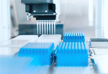
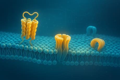
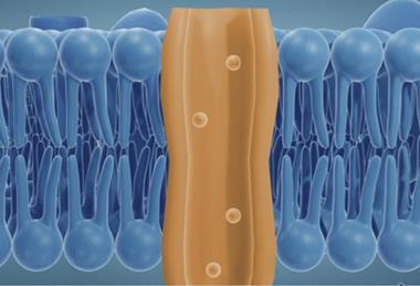
From expert task to lab routine – Membrane protein purification and analysis simplified
A new way to tackle the most challenging drug targets: Discover a fully automated, detergent-free purification workflow that isolates membrane proteins in their native 3D structure in as little as three hours and with minimal hands-on time. Learn how to perform subsequent digital miniaturization and qPCR-based stability analysis with compact solutions that integrate seamlessly into your lab.
Where NAMs stand in early drug discovery: an expert discussion
From expert task to lab routine – Membrane protein purification and analysis simplified
On-demand Webinars
- Previous
- Next


Whitepapers
- Previous
- Next