In this Q&A, Keith Murphy, CEO of Organovo, discusses how the shift from animal models to 3D human tissues can transform the preclinical space. Among his many insights, he details the limitations of animal models, how Organovo’s 3D human tissue models facilitate a deeper understanding of genetic expression within specific diseases, and how widespread adoption of these models will enable rapid drug target identification, reduce cost and decrease toxicity.

What specific limitations in mirroring diseases in humans are commonly observed when using animal models for preclinical research?
The standard animal model paradigm for drug discovery does not translate well to effects in patients for a number of reasons. For one, animal models require disease induction, either genetic or surgical modifications, diet, or application of a disease inducing agent or toxin, yet this is nothing like how diseases progress in humans, which typically take years to develop. Furthermore, there are genetic differences in animals compared to humans; for example, there is a 15 percent difference in mouse and human protein coding sequences, and, when multiplied by 140,000 genes, this creates vast differences. Another important factor is that there are gene expression differences between animals and humans, such as chromosomes, enhancers and gene promoter sequences that are significantly rearranged when comparing animals to humans, which leads to differences in expression levels for many genes. In fact, human genetic variation is relatively high, while inbred mouse strains have lower genetic variations as a population as they have been inbred for hundreds of years to be as homozygous as possible. Lastly, it’s important to note that the experience of the individual patient is captured epigenetically, including the disease profile in many cases, but animal models simply don’t have the disease history to show the right profiles.
In summary, all these factors can affect the response to drugs and drug target manipulation. While animal models have been extremely successful despite these challenges, the worsening clinical failure rate [note: 88 percent from 2000-2010; 92 percent from 2010-2020] for currently targeted diseases suggests that animal models helped find the lower hanging fruit and we may need to consider a new approach for the remaining, more complex, diseases.
How do three-dimensional (3D) human tissue models accurately replicate cellular interactions within living tissue systems compared to traditional cell or tissue cultures?
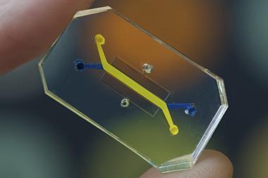
The Organovo 3D human tissue models replicate cellular interactions in a number of ways. By creating models from primary cells – those cells that have the specific disease profile when in the patient - the challenge is no longer to “create” the disease from scratch, but to keep it active in the same cells as they form a new tissue. Much discovery work is done in cell lines, which is convenient and rapid because they live forever, but they are not representative of typical human tissue. Our cells are fully differentiated, non-tumour, primary cells, which have a more natural growth pattern and gene expression profile, and the disease signature is often carried over epigenetically. Furthermore, our three-dimensional culture system allows for the formation of a structure in part developed by the input cells that facilitates natural cell-cell connections, more natural biochemical and mechanical cell-cell signalling. By using multiple cell types, we can allow cells to find their normal partners during formation, interacting directly as they would in the native tissue to ensure the best response. We are targeting human primary cells that already have the disease and carry the signals from their time in the diseased state from the patient. By analysing these signals and using them to develop drugs, we are able to enhance the chances that those drugs will be effective in the clinic.
Compared to conventional models, could you explain how Organovo's 3D tissue models facilitate a deeper understanding of genetic expression within specific diseases, and how this insight might transform the development of targeted therapies?
Once created, our 3D human tissue models can be manipulated and worked with in the lab. Since they are made from primary cells, there is a high comparability to clinical gene expression profiles and we are able to conduct superior experimentation comparable to our ability in the clinic. As an example, we can see gene expression profile responses to interventions that are directly relevant to the human biology.
How does Organovo's use of 3D tissue technology to model inflammatory bowel diseases, particularly in the case of FXR314, pave the way for more effective drug discovery and potential therapies for a range of diseases beyond ulcerative colitis?
Using our 3D human IBD model, we have at our disposal a powerful tool to increase the probability of success of our therapeutic candidates, including FXR314, which has already demonstrated proof of concept. FXR314 has demonstrated activity in the tissue and not simply as a global anti-inflammatory blockade, which points to our models having the ability to locate tissue-specific autoimmune disease targets for other diseases as well.
What potential impact do you foresee the widespread adoption of 3D tissue models having on the future of drug discovery and development within the biopharmaceutical industry?
We believe that widespread adoption of 3D human tissue models will allow us to identify effective drug targets much more rapidly and enable us to pursue them with increased vigour without waiting for long-term animal model data. Furthermore, we believe that an increased understanding of the predictive value of 3D tissue models will alter and shorten the regulatory pathway for new drugs and potentially reduce the overall cost, which could in turn bring down drug prices. We foresee that the use of animal models to assess efficacy and eventually toxicity will decrease, reducing the number of animals sacrificed for research. Also, adoption of 3D human tissue models will help reduce the number of failed clinical trials because ineffective drugs are weeded out pre-clinically. Lastly, by using 3D human tissue models, patients in clinical trials will be exposed to fewer ineffective drugs, reducing their risk of side effects without any benefit.
Author bio
 Keith Murphy, CEO of Organovo
Keith Murphy is currently CEO of Organovo and a serial entrepreneur and investor in biotech. Mr Murphy founded Organovo in 2007 and led all company operations until 2017, when he co-founded Viscient Biosciences. He co-invented the NovoGen MMX bioprinter platform and grew Organovo through early investments and pharma corporate partnerships.
Prior to Organovo, Mr. Murphy spent 10 years at Amgen in roles of increasing responsibility, including four years as the Global Operations Leader of denosumab, now marketed as Prolia & Xgeva ($4B+ annual sales). Prior to Amgen, he played a key role at Alkermes in the successful development of once-monthly human growth hormone (hGH) in partnership with Genentech.
He holds a B.S. in Chemical Engineering from the Massachusetts Institute of Technology and is an alumnus of the UCLA Anderson School of Management. Mr Murphy serves as co-chair of the Board of No Patient Left Behind, and on the Board of Directors of the California Life Sciences Association, the state’s life science industry public policy, advocacy and business leadership organisation.