Calcilytics as potential novel therapeutic treatments to halt Alzheimer’s disease
Posted: 7 September 2017 | Dr Anna Chiarini, Dr Ilaria Dal Prà, Prof Ubaldo Armato | No comments yet
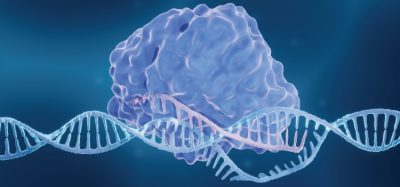
Alzheimer’s disease (AD) is the most prevalent form of dementia.1 The common (>95% of cases) slow-developing form of the ailment is known as late-onset AD (LOAD) or sporadic AD (SAD). The rare (<5% of cases), faster-developing form of the disease is early-onset AD (EOAD) or familial AD (FAD), the symptoms of which manifest at an unusually early age (under 60). These patients carry a mutant gene for the β-secretase 1 (BACE1), or more often the presenilin (PSEN1 or PSEN2), components of the γ-secretase complex causing the overproduction of amyloid-β42 oligomers (Aβ42-os).2
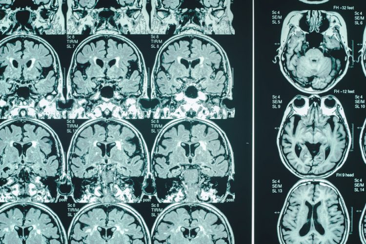
SAD affects millions of people in the Western world.3 Its prevalence increases, reaching around 50% by 85 years of age but has been imperceptibly developing for decades with a slow intra-brain accumulation: first of toxic Aβ42-os, and later of hyperphosphorylated Tau protein oligomers (p-Tau-os). These produce micro-ischemic foci, neuroinflammation, synaptic loss, and neuronal death starting within the entorhinal cortex layer II and the hippocampus and subsequently spreading to cognition-linked upper cortical areas. Eventually, SAD emerges clinically with progressive memory failure.4-6
A lot of effort has been invested in trying to stop or reverse the neuronal damage inflicted by SAD. Hitherto, treating human SAD with drugs or otherwise has met with total failure.7,8 In fact, SAD is a complex human disorder that is only partially modelled in rodents, and examining the effect of potential drugs in animal models has had a low predictive power for treatment of SAD patients. To bridge the gap between animal studies and clinical trials, cortical normo-functioning (untransformed) adult human astrocytes (NAHAs) and postnatal human HCN-1A neurons have been used as a preclinical paradigm. This has recently disclosed exciting evidence of a potential treatment for SAD based on the following discoveries…
- The Ca2+-sensing receptor (CaSR) signalling pathologically activated by exogenous Aβ42-os plays crucial roles in the neuronal damage inflicted by SAD
- Highly specific allosteric CaSR antagonists named ‘calcilytics’ are able to suppress the deathly effects due to the Ab-osCaSR signalling.9-1
CaSR background
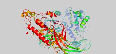
CaSR is a member of the family C of G-protein (heterotrimeric guanine nucleotide–binding protein)-coupled receptors (GPCRs) or seven-transmembrane receptors that play a primary role in the regulation of various physiological and pathophysiological processes, making the GPCR superfamily a major target for therapeutic intervention.12 CaSRs can form homodimers (CaSR/CaSR) or heterodimers (CaSR/mGluR). The receptor detects and responds to changes in [Ca2+]e to maintain the ion’s physiological concentration (1.1–1.3mM). However, the CaSR is a relatively non-selective cationic receptor. Among the many CaSR ligands are di- and trivalent cations (Mg2+, Ba2+, Cd2+, Co2+, Fe2+, Gd3+, Ni2+, Pb2+), aromatic L-α-amino acids (L-α-Phe, L-α-Trp, L-α-Tyr, and L-α-His), aminoglycoside antibiotics (neomycin, gentamicin, tobramicin), polyamines, and last but not least, Aβ42-os.13
CaSR and AD
In fact, most important in the sphere of AD is the ability of Aβ peptides to bind and activate the CaSR. Indeed, the toxic Aβ42-os and fibrillar Aβ42 aggregates, like the cation and polyamine CaSR ligands, have regular arrays of positive charges and can bind the CaSR. Recent studies9,13-15 have shown that exogenous Aβ25-35-os and Aβ42-os bind with a high specificity the plasmalemmal CaSRs of NAHAs and HCN-1A neurons, thereby activating a pathological Aβ-os·CaSR signalling that triggers a set of neurotoxic processes:
- An overexpression of the CaSR that sensitises neurons and astrocytes to toxic Aβ42-os.
- An intracellular build-up of endogenous Aβ42-os in both NAHAs and HCN-1A neurons#b# enhanced by the reduced activities of the proteasome and various Aβ-cleaving enzymes.
- An increased secretion of the accumulating endogenous Aβ42-os from both NAHAs and HCN-1A neurons, which pushes the extracellular Aβ42/Aβ40 ratio into the frankly neurotoxic range. Moreover, the oversecreted Aβ-os specifically bind and activate the neurons’ and astrocytes’ overexpressed CaSRs, thereby leading a set of feed-forward continual vicious loops in both cell types promoting a progressive extracellular accumulation and diffusion of cytotoxic Aβ-os.
- An increased activity of GSK-3β, ie, one of the main Tau protein kinases, which produces phospho-Taues subsequently aggregating as p-Tau-os.16
- An increase in the intracellular p-Tau-os levels, which are next released extracellularly inside exosomes over basal levels from NAHAs. 16
- A surplus production/release of nitric oxide (NO) from NAHAs via a MEK/ERK1/2-dependent activation of the induced GTP cyclohydrolase-1 (GCH1) producing a cofactor, tetrahydrobiopterin (or BH4) that mediates the dimerisation and activation of the co-induced nitric oxide (NO) synthase-2 (NOS-2).9
- An excess production/secretion of VEGF-A (mostly VEGF-A165) from NAHAs prompted by HIF-1a stabilisation and nuclear translocation; note that at high concentrations VEGF-A is neurotoxic.14
- A slow yet progressive death of the human cortical HCN-1A neurons in vitro like the one underlying the growing loss of cognitive functions in vivo.9 Conversely, the Aβ-os-exposed NAHAs survive and keep producing, releasing, and spreading neuron-killing Aβ42-os, NO, and VEGF-A165.
Therefore, these findings reveal how crucial and critical the role played by Aβ-os·CaSR signalling can be in the progressive spreading of human SAD’s neuropathology within the cognition-linked cerebro-cortical areas.
It is worth recalling here that all kinds of cells resident in the human central nervous system significantly express the CaSR: hence, once exposed to exogenous Aβ-os they can be recruited via Aβ-os•CaSR signalling to oversecrete further amounts of Aβ-os, which can start new cycles of self-inducing and self-sustaining Aβ-os spreading, thereby promoting AD progression. For this reason, CaSR has emerged in recent years as an attractive target for drug development to treat AD.
CaSR as drug target
It should be clear, as the above-mentioned observations indicate, that stopping the pathological Aβ·CaSR signalling in human cortical astrocytes and neurons should be the first therapeutic target in order to halt AD.
Synthetic CaSR antagonists, or calcilytics, represent a set of valuable tools for investigating the CaSR signalling pathways, for manipulating and modulating CaSR’s function, and for treating disorders involving the CaSR. Chemically, calcilytic compounds are quinazolinones like ATF936 and AXT914, or small organic molecules based on amino-alcohol structures like NPS 2143, Ronacaleret, Calhex 231, NPSP795, JTT-305, which act as negative allosteric modulators of the CaSR.17,18 They right-shift the [Ca2+]e response curve, decreasing the sensitivity of the CaSR to [Ca2+]e values and thus increasing the EC50 for [Ca2+]e. Several calcilytics have been evaluated as therapeutics for postmenopausal osteoporosis, but clinical development was abandoned because they were found to be ineffective. Currently, they have been repurposed for new therapeutic indications like hypoparathyroidism, and in autosomal dominant hypocalcaemia (ADH), as well as the treatment of cancers where CaSR-mediated signalling is involved in the establishment of bone metastases of prostate and breast cancer cells.19,20 Other recent translational suggestions for calcilytics include asthma attacks, pulmonary artery idiopathic hypertension, and stroke.21-23
Focusing on AD, it has been demonstrated that Aβ-os·CaSR noxious signalling can be effectively and completely antagonised by highly selective CaSR allosteric antagonists17,18 – indeed a most important therapeutic asset.9,11,14,16 Calcilytics like NPS 2143 and NPS 89686 fully suppress all the mentioned effects of Aβ-os·CaSR signalling, including the excess secretion and diffusion of endogenously produced Aβ-os – the first neurotoxic drivers of AD according to the ‘amyloid cascade hypothesis’ – from exogenous Aβ-os-exposed human neurons and astrocytes cultured in vitro, and blocks the propagation of toxic p-Tau-os and likely the consequent tauopathy.9,11,14,16 Hence, calcilytics would halt or attenuate the Aβ-os-elicited amyloidosis, tauopathy, synaptotoxicity, angiopathy, neuroinflammation, myelin sheaths degeneration and, most of all, the cortical neurons death and consequently, the otherwise inexorable SAD progression in vivo.
Adding to the appeal of CaSR as a drug target to treat AD is the recent discovery that NPS 2143 has the potential to restore the physiological release of the soluble non-amyloidogenic form of the beta-amyloid precursor protein α (sAPPα) that is known to exert neuroprotective and neurotrophic effects.11 These findings gained from preclinical models made of cultured human untransformed neural cells indicate that, by effectively antagonising the amyloidogenic processing of APP promoted by Aβs•CaSR signalling, in vivo administered calcilytics would preserve the shedding of neurotrophic sAPPα from the plasma membrane of astrocytes and likely neurons. Therefore, calcilytics would keep the in vivo human neurons alive and functioning as they do in vitro, safeguarding the memory and cognition of the patients.
Conclusion
All these preclinical findings have disclosed molecular mechanisms that likely play a role in SAD’s onset and progression and raise the enticing prospect that pathological Ab·CaSR signalling simultaneously triggers both the Ab-mediated and the p-Tau-mediated neurotoxic mechanisms driving SAD neuropathology. The exciting focus of these discoveries is that calcilytics like NPS 2143 can fully suppress all the neurotoxic effects that Ab·CaSR signalling elicits. Hence, by antagonising the Aβs•CaSR signalling, calcilytics hold the potential to serve as a novel therapy for SAD and to act as useful drugs at the earliest SAD stage detectable in order to hinder intrabrain Aβ-os and pTau-os spreading, and hence SAD progression.
Author Biographies
 Dr Anna Chiarini is Assistant Professor at Verona University Medical School, Italy. After obtaining a BSc in Biological Sciences from Milan University (Italy), she gained her PhD in Chronic and Degenerative Diseases from Verona University. Her scientific interests are currently focused on Alzheimer’s Disease pathogenesis using omics strategies and cellular and biochemical assays.
 Prof Ubaldo Armato is Senior Investigator of the Histology & Embryology Unit, Verona University Medical School (Italy). He majored in Medicine and Surgery at the University of Padua, Italy. His research interests encompass the proteomic and biochemical mechanisms of Alzheimer’s disease pathogenesis and in vitro and in vivo testing of innovative biomaterials for tissue regeneration.
 Dr Ilaria Dal Prà graduated in Biological Sciences from Milan University (Italy) and gained a PhD in Molecular and Cellular Biology and Pathology from Padua University. She is Assistant Professor in the Histology and Embryology Unit at Verona University Medical School, and works on Alzheimer’s disease pathogenesis. She expertly uses culture normal adult human astrocytes and neurons to investigate the disease molecular mechanisms.
References
- Tarawneh R, Holtzman DM. The clinical problem of symptomatic Alzheimer disease and mild cognitive impairment. Cold Spring Harb Perspect Med. 2012 May;2(5):a006148. doi: 10.1101/cshperspect.a006148. Review.
- Andrew RJ, Kellett KA, Thinakaran G, Hooper NM. A Greek Tragedy: the growing complexity of Alzheimer amyloid precursor protein proteolysis. J Biol Chem. 2016 Sep;291(37):19235-44.
- Zhao LN, Lu L, Chew LY, Mu Y. Alzheimer’s disease-a panorama glimpse. Int J Mol 2014 Jul;15(7):12631-50.
- Marchesi VT. Alzheimer’s dementia begins as a disease of small blood vessels, damaged by oxidative-induced inflammation and dysregulated amyloid metabolism: implications for early detection and therapy. FASEB J. 2011 Jan;25(1):5-13.
- Khan UA, Liu L, Provenzano FA, Berman DE, Profaci CP, Sloan R, et al. Molecular drivers and cortical spread of lateral entorhinal cortex dysfunction in preclinical Alzheimer’s disease. Nat Neurosci. 2014 Feb;17(2):304-11.
- Viola KL, Klein WL.Amyloid β oligomers in Alzheimer’s disease pathogenesis, treatment, and diagnosis. Acta Neuropathol 2015 Feb;129(2):183-206.
- Berk C, Sabbagh MN. Successes and failures for drugs in late-stage development for Alzheimer’s disease. Drugs Aging. 2013 Oct;30(10):783-92. Erratum in Drugs Aging 2014 Jan;31(1):81.
- Rosenblum WI. Why Alzheimer trials fail: removing soluble oligomeric beta amyloid is essential, inconsistent, and difficult. Neurobiol Aging 2014 May;35(5):969-74.
- Armato U, Chiarini A, Chakravarthy B, Chioffi F, Pacchiana R, Colarusso E, et al. Calcium-sensing receptor antagonist (calcilytic) NPS 2143 specifically blocks the increased secretion of endogenous Aβ42 prompted by exogenous fibrillary or soluble Aβ25-35 in human cortical astrocytes and neurons-therapeutic relevance to Alzheimer’s disease. Biochim Biophys Acta. 2013 Oct;1832(10):1634-52.
- Chiarini A, Armato U, Liu D, Dal Prà I. Calcium-Sensing Receptors of Human Neural Cells Play Crucial Roles in Alzheimer’s Disease. Front Physiol. 2016 Apr 26;7:134. doi: 10.3389/fphys.2016.00134. eCollection 2016.
- Chiarini A, Armato U, Liu D, Dal Prà I. Calcium-Sensing Receptor AntagonistNPS 2143 Restores Amyloid Precursor Protein Physiological Non-Amyloidogenic Processing in Aβ-Exposed Adult Human Astrocytes. Sci Rep. 2017 Apr 28;7(1):1277. doi: 10.1038/s41598-017-01215-3.
- Salon JA, Lodowski DT, Palczewski K. The significance of G protein-coupled receptor crystallography for drug discovery. Pharmacol Rev. 2011Dec;63(4):901-37. doi: 10.1124/pr.110.003350.
- Dal Prà I, Chiarini A, Pacchiana R, Gardenal E, Chakravarthy B, Whitfield JF, et al. Calcium-Sensing Receptors of Human Astrocyte-Neuron Teams: Amyloid-β-Driven Mediators and Therapeutic Targets of Alzheimer’s Disease. Curr 2014 Jul;12(4):353-64. doi: 10.2174/1570159X12666140828214701.
- Dal Prà I, Armato U, Chioffi F, Pacchiana R, Whitfield JF, Chakravarthy B, et al. The Aβ peptides-activated calcium-sensing receptor stimulates the production and secretion of vascular endothelial growth factor-A by normoxic adult human cortical astrocytes. Neuromolecular Med. 2014 Dec;16(4):645-57. doi: 10.1007/s12017-014-8315-9.
- Dal Prà I, Chiarini A, Gui L, Chakravarthy B, Pacchiana R, Gardenal E, et al. Do astrocytes collaborate with neurons in spreading the “infectious” aβ and Tau drivers of Alzheimer’s disease? Neuroscientist. 2015Feb;21(1):9-29. doi: 10.1177/1073858414529828.
- Chiarini A, Armato U, Gardenal E, Gui L, Dal Prà I. Amyloid β-Exposed Human Astrocytes Overproduce Phospho-Tau and Overrelease It within Exosomes, Effects Suppressed by Calcilytic NPS 2143-Further Implications for Alzheimer’s Therapy. Front Neurosci. 2017 Apr 20;11:217. doi: 10.3389/fnins.2017.00217.
- Nemeth EF and Goodman WG. Calcimimetic and calcilytic drugs: feats, flops, and futures. Calcif Tissue Int 2016 Apr;98(4):341-58. doi: 10.1007/s00223-015-0052-z.
- Nemeth EF. Allosteric modulators of the extracellular calcium receptor. Drug Discov Today Technol. 2013 Summer;10(2):e277-84. doi:10.1016/j.ddtec.2012.11.002.
- Hannan FM, Walls GV, Babinskis VN, Nesbit MA, Kallay E, Hough TA, et al. The Calcilytic Agent NPS 2143 Rectifies Hypocalcemia in a Mouse Model with an Activating Calcium-Sensing Receptor (CaSR) Mutation: Relevance to Autosomal Dominant Hypocalcemia Type 1 (ADH1). Endocrinology. 2015 Sep;156(9):3114-21. doi: 10.1210/en.2015-1269.
- Singh N, Promkan M, Liu G, Varani J, Chakrabarty S. Role of calcium sensing receptor (CaSR) in tumorigenesis. Best Pract Res Clin Endocrinol Metab. 2013 Jun;27(3):455-63. doi: 10.1016/j.beem.2013.04.001.
- Yarova PL, Stewart AL, Sathish V, Britt RD Jr, Thompson MA, P Lowe AP, et al. Calcium-sensing receptor antagonists abrogate airway hyperresponsiveness and inflammation in allergic asthma. Sci Transl Med. 2015 Apr 22;7(284):284ra60. doi:1126/scitranslmed.aaa0282.
- Yamamura A, Ohara N, Tsukamoto K. Inhibition of Excessive Cell Proliferation by Calcilytics in Idiopathic Pulmonary Arterial Hypertension. PLoS One. 2015 Sep 16;10(9):e0138384. doe: 10.1371/journal.pone.0138384.
- Kim JY, Ho H, Kim N, Liu J, Tu CL, Yenari MA, et al. Calcium-sensing receptor (CaSR) as a novel target for ischemic neuroprotection. Ann CLIN Trans Neurol. 2014 Nov;1(11):851-66. doe: 10.1002/acn3.118.
Related topics
Biomarkers, Drug Targets, Genomics
Related conditions
Alzheimer’s disease
Related organisations
University of Verona
Related people
Anna Chiarini, Dr Ilaria Dal Prà, Prof Ubaldo Armato